

Definitions
Definitions Based on Systemic inflammatory Response Syndrome (SIRS) Criteria
- Systemic Inflammatory Response Syndrome (SIRS) Criteria: any of the following
- Fever (Temperature >100.9° F) or Hypothermia (Temperature <96.8° F) (see Fever)
- Leukocytosis (White Blood Cell >12k) or Leukopenia (WBC <4k) or Bandemia (Bands >10%) (see Leukocytosis)
- Tachycardia (Heart Rate >90 bpm) (see Sinus Tachycardia)
- Tachypnea (Respiratory Rate >20 breaths/min) (see Tachypnea)
- Sepsis: including both of the following
- Suspected or Possible Source of Infection
- ≥2 Systemic Inflammatory Response Syndrome (SIRS) Criteria
- Severe Sepsis: including both of the following
- Sepsis
- Organ Dysfunction Criteria
- Hypotension (see Hypotension)
- Hypoxemia/Respiratory Failure (see Hypoxemia and Respiratory Failure)
- Oliguria/Acute Kidney Injury (AKI) (see Acute Kidney Injury)
- Hepatic Dysfunction
- Thrombocytopenia (see Thrombocytopenia)
- Coagulopathy (see Coagulopathy)
- Lactic Acidosis (see Lactic Acidosis)
- Septic Shock: including both of the following
- Severe Sepsis
- Hypotension Persisting in the Hour After the Intravenous Fluid Bolus as Evidenced By Either of the Following (see Hypotension)
- Systolic Blood Pressure <90 or Mean Arterial Pressure <65 or a Systolic Blood Pressure Decrease of >40 mm Hg
- Tissue Hypoperfusion Present with Initial Serum Lactate Level ≥4 mmol/L
Third International Consensus Definitions for Sepsis and Septic Shock (Sepsis-3: Society of Critical Care Medicine and European Society of Intensive Care Medicine) (JAMA, 2016) [MEDLINE]
- Sepsis: life-threatening organ dysfunction caused by dysregulated host response to infection
- Organ Dysfunction: an infection-related acute change in sequential organ failure assessment (SOFA) score ≥2 pts
- Sepsis Mortality Rate: approximate 10% mortality rate
- Septic Shock: sepsis with persistent vasopressor-dependent hypotension (to maintain MAP ≥65 mm Hg) and serum lactate level >2 mmol/L despite adequate intravenous volume resuscitation
- Septic Shock Mortality Rate: >40% mortality rate

Epidemiology
Incidence
- Incidence of Septic Shock is Increasing
- Retrospective Study of Central Venous Catheter Use in Septic Shock Using Data from the Nationwide Inpatient Sample (NIS), 1998-2009 (Crit Care Med, 2013) [MEDLINE]: n = 203,481 admitted through the ED with septic shock
- From 1998 to 2009, US Adult Population-Adjusted Rates of Septic Shock Increased from 12.6 Cases Per 100k to 78 Cases Per 100k
- Systematic Review of the Global Incidence and Mortality of Sepsis (Am J Respir Crit Care Med, 2016) [MEDLINE]: studies from 1979 to 2015
- In Articles Restricted to the Last 10 Years
- Sepsis Incidence Rate: 437 Cases Per 100k
- Severe Sepsis Incidence Rate: 270 Cases Per 100k
- In Articles Restricted to the Last 10 Years
- Sepsis In-Hospital Mortality Rate: 17%
- Severe Sepsis In-Hospital Mortality Rate: 26%
- In Articles Restricted to the Last 10 Years
- Study of the Incidence and Trends of Sepsis in US Hospitals Using Clinical vs Claims Data (2009-2014) (JAMA, 2017) [MEDLINE]
- In Clinical Data from 409 Hospitals, Sepsis was Present in 6% of Adult Admissions, While in Claims-Based Data, Neither the Incidence of Sepsis Nor the Combined Outcome or Death or Discharge to Hospice Changed Significantly from 2009-2014
- Findings Suggest that Electronic Health Record-Based Clinical Data Provide More Objective Estimates than Claims-Based Data for Sepsis Surveillance
- Study of Septic Shock Incidence and Mortality Rate in United States Academic Medical Centers Using Clinical Data (Chest, 2017) [MEDLINE]: of 6.5 million adult hospitalizations, 99,312 (1.5%) were flagged by clinical criteria, 82,350 (1.3%) by ICD-9 codes, and 44,651 (0.7%) by both
- Sensitivity for Clinical Criteria was Higher than Claims (74.8% vs 48.3%), Whereas Positive Predictive Value was Comparable (83% vs 89%)
- Septic Shock Incidence (Based on Clinical Criteria) Increased from 12.8 to 18.6 Cases Per 1,000 Hospitalizations (Average, 4.9% Increase Per Year; 95% CI, 4.0%-5.9%), While the Mortality Rate Decreased from 54.9% to 50.7% (Average, 0.6% Decline Per Year; 95% CI, 0.4%-0.8%)
- In Contrast, Septic Shock Incidence (Based on ICD-9 Codes) Increased from 6.7 to 19.3 Per 1,000 Hospitalizations (19.8% Increase Per Year; 95% CI, 16.6%-20.9%), While the Mortality Rate Decreased from 48.3% to 39.3% (1.2% Decline Per Year; 95% CI, 0.9%-1.6%)
- Clinical Surveillance Definition (Based on Concurrent Vasopressors, Blood Cultures, and Antibiotics Accurately Identified Septic Shock Hospitalizations
- Incidence of Patients Receiving Treatment for Septic Shock Has Increased and Mortality Rates Have Decreased (But Less Dramatically than Estimated on the Basis of ICD-9 Billing Codes)
- Retrospective Study of Central Venous Catheter Use in Septic Shock Using Data from the Nationwide Inpatient Sample (NIS), 1998-2009 (Crit Care Med, 2013) [MEDLINE]: n = 203,481 admitted through the ED with septic shock
Seasonality
- Incidence of Sepsis and Severe Sepsis are Highest in Winter Months (Predominantly Associated with an Increased Risk of Respiratory Infections) (Crit Care Med, 2007) [MEDLINE]: retrospective study
- Seasonal Incidence Rate of Sepsis Increased from 41.7 Cases Per 100k in the Fall to 48.6 Cases Per 100k in the Winter
- Seasonal Incidence Rate of Severe Sepsis Increased from 13.0 Cases Per 100k in the Fall to 15.3 Cases Per 100k in the Winter
- Seasonal Changes in the Incidence of Sepsis Varied According to Geographic Region: greatest seasonal change in sepsis rates occurred in the Northeast (+30%)
- Sepsis Case-Fatality Rates Were 13% Greater in the Winter, as Compared to the Summer, Despite Similar Severity of Illness
Disease Severity
- Sepsis Severity is Increasing
- Rate of Severe Sepsis Hospitalization Doubled from 1993 to 2003 (Crit Care Med, 2007) [MEDLINE] (Crit Care Med, 2013) [MEDLINE]
- Mortality from Severe Sepsis Also Increased Significantly During This Period
- However, Case Fatality Rates Decreased During This Period
- Study of Septic Shock Incidence and Mortality Rate in United States Academic Medical Centers Using Clinical Data (Chest, 2017) [MEDLINE]: of 6.5 million adult hospitalizations, 99,312 (1.5%) were flagged by clinical criteria, 82,350 (1.3%) by ICD-9 codes, and 44,651 (0.7%) by both
- Incidence of Patients Receiving Treatment for Septic Shock Has Increased and Mortality Rates Have Decreased (But Less Dramatically than Estimated on the Basis of ICD-9 Billing Codes)
- Rate of Severe Sepsis Hospitalization Doubled from 1993 to 2003 (Crit Care Med, 2007) [MEDLINE] (Crit Care Med, 2013) [MEDLINE]
Risk Factors
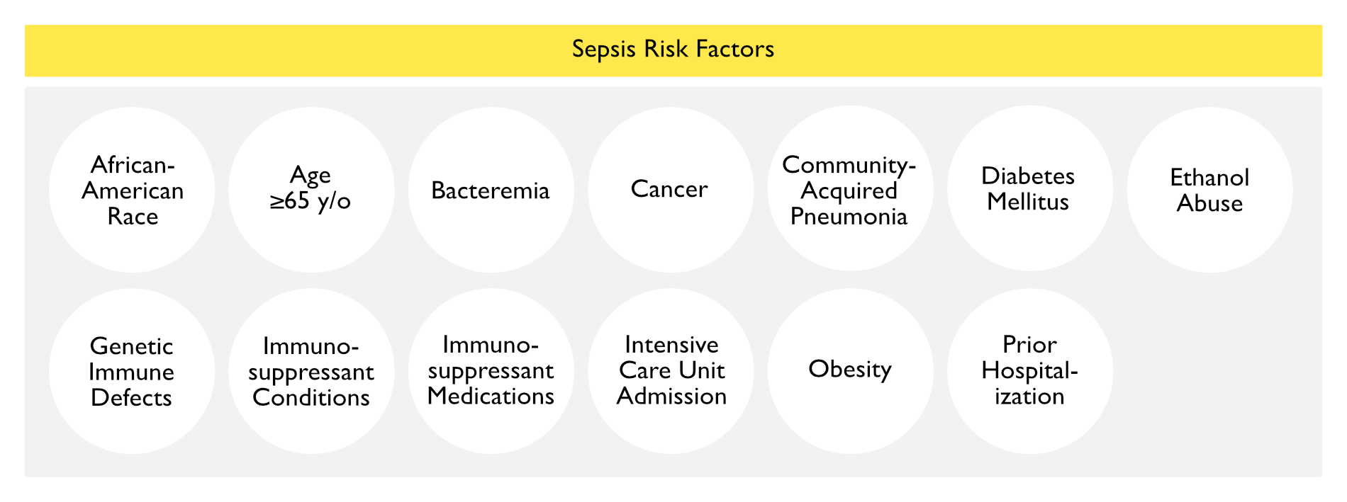
- African-American Race
- Sepsis Incidence is Highest in African-American Males (NEJM, 2003) [MEDLINE]
- Age ≥65 y/o
- Up to 65% of Patients Who Develop Severe Sepsis in the US are ≥65 y/o (Clin Infect Dis, 2005) [MEDLINE]
- Age Independently Increases Both the Risk of Sepsis and the Mortality Rate Associated with Sepsis (Crit Care Med, 2006) [MEDLINE]
- Elderly Patients Account for 12% of the US Population, But 65% of Sepsis Cases (Relative Risk 13.1, as Compared to Younger Patients)
- Elderly Patients are More Likely to Have Gram-Negative Infections, Particularly in Association with Pneumonia (Relative Risk 1.66, as Compared to Younger Patients)
- Case-Fatality Rates Increase Linearly with Age
- Age is an Independent Predictor of Mortality (Odds Ratio 2.26)
- Elderly Sepsis Patients Died Earlier During Hospitalization
- Elderly Survivors of Sepsis were More Likely to Be Discharged to a Non-Acute Health Care Facility
- Bacteremia (see Bacteremia)
- Bacteremia is Frequently Associated with Sepsis, with 95% of Bacteremic Patients Meeting SIRS Criteria (QJM, 1996) [MEDLINE]
- Cancer
- Cancer Increases the Risk of Hospitalization with Severe Sepsis (Relative Risk 3.96) (Crit Care, 2004) [MEDLINE]
- As Compared with the General Population, Cancer Patients are Much More Likely to Be Hospitalized (Relative Risk, 2.77; 95% CI, 2.77-2.78) and to Be Hospitalized with Severe Sepsis (Relative Risk, 3.96; 95% CI, 3.94-3.99)
- Patients with a History of Cancer are at Increased Risk for Acquiring and Dying from Sepsis, as Compared to the General Population (Chest, 2006) [MEDLINE]: however, incidence and fatality rates are decreasing over time
- Cancer Increases the Risk of Hospitalization with Severe Sepsis (Relative Risk 3.96) (Crit Care, 2004) [MEDLINE]
- Community-Acquired Pneumonia (CAP) (see Community-Acquired Pneumonia)
- Diabetes Mellitus (DM) (see Diabetes Mellitus)
- Diabetes Mellitus Impairs Immune System Function and Increases the Risk of Infection (and Sepsis) (Diabetes Care, 2003) [MEDLINE]
- Diabetes Mellitus is Not Associated with Changes in Sepsis Presentation or 90-Day Mortality Rate (Crit Care, 2016) [MEDLINE]
- Patients with Type 2 Diabetes Taking Sodium-Glucose Cotransporter-2 (SGLT2) Inhibitors are at Decreased Risk for Pneumonia and Sepsis, as Compared to Those Taking Dipeptidyl Peptidase-4 (DPP-4) Inhibitors (Diabetes Metab, 2022) [MEDLINE] (see Sodium-Glucose Cotransporter-2 Inhibitors and Dipeptidyl Peptidase-4 Inhibitors)
- Ethanol Abuse (see Ethanol
- Genetic Factors
- Various Genetic Defects (Antibody Synthesis Defects, T-Cell Deficiency, Phagocyte Deficiency, Natural Killer Cell Deficiency, Complement Deficiency, or Impaired Innate Immunity Pathogen Pattern Recognition Receptors) Have Been Demonstrated to Increase the Risk of Sepsis (N Engl J Med, 2011) [MEDLINE]
- Defects in Toll-Like Receptors (TLR’s): defects/deficiencies have been identified in TLR3, TLR5, MyD88, IRAK-4, UNC93B
- Defects in C-Type Lectin Receptors (CLR’s): defects/deficiencies have been identified in Dectin-1, CARD-9, Mannose-Binding Lectin
- Defects in Nucleotide-Binding Oligomerization Domain (NOD) Leucine-Rich-Repeat Containing Receptors (NLR’s): defects/deficiencies have been identified in NOD2, NLRP3
- Complement Deficiency
- T-Cell Defects
- Phagocyte Defects
- Various Genetic Defects (Antibody Synthesis Defects, T-Cell Deficiency, Phagocyte Deficiency, Natural Killer Cell Deficiency, Complement Deficiency, or Impaired Innate Immunity Pathogen Pattern Recognition Receptors) Have Been Demonstrated to Increase the Risk of Sepsis (N Engl J Med, 2011) [MEDLINE]
- Immunosuppression
- Immunosuppressing Conditions
- Acquired Immunodeficiency Syndrome (AIDS) (see Human Immunodeficiency Virus)
- Asplenia (see Asplenia)
- Cancer
- Chronic Kidney Disease (CKD) (see Chronic Kidney Disease)
- Liver Disease (see End-Stage Liver Disease) (Chest, 2003) [MEDLINE]
- Corticosteroids (see Corticosteroids)
- United States Population-Based Retrospective Cohort Study of the Risks of Short-Term Corticosteroid Use in Adults (BMJ, 2017) [MEDLINE]
- One in Five American Adults in a Commercially-Insured Plan were Given at Least One Outpatient Short-Term Corticosteroid Course During the Three Year Study (2012-2014): mostly for upper respiratory tract infections, spinal conditions, and allergies
- Within 30 Days of Initiation, Short-Term Use of Corticosteroids Increased the Risk of Sepsis (Incidence Rate Ratio 5.30, 95% CI: 3.80-7.41), Venous Thromboembolism (Incidence Rate Ratio 3.33, 95% CI: 2.78 to 3.99), and Fractures (Incidence Rate Ratio 1.87, 95% CI: 1.69 to 2.07): increased risk persisted at prednisone equivalent doses of <20 mg/day (incidence rate ratio 4.02 for sepsis, 3.61 for venous thromboembolism, and 1.83 for fracture)
- United States Population-Based Retrospective Cohort Study of the Risks of Short-Term Corticosteroid Use in Adults (BMJ, 2017) [MEDLINE]
- Other Immunosuppressant Medications
- Cyclosporine A (see Cyclosporine A)
- Mycophenolate Mofetil (Cellcept) (see Mycophenolate Mofetil)
- Tacrolimus (see Tacrolimus)
- Immunosuppressing Conditions
- Intensive Care Unit (ICU) Admission
- Intensive Care Unit Admission is a Risk Factor for Infection with Drug-Resistant Organisms, Which May Lead to Sepsis (JAMA, 1995) [MEDLINE]
- Obesity (see Obesity)
- Obesity is Believed to Impair Immune System Function, Increasing the Risk of Infections Which May Lead to Sepsis (Especially Aspiration Pneumonia During Hospitalization, Community-Acquired Pneumonia, Biliary Tract Disease, Skin Infections) (Lancet Infect Dis, 2006) [MEDLINE]
- Prior Hospitalization
- Hospitalization Alters the Microbiome (Especially in Patients Treated with Antibiotics), Increasing the Risk of Sepsis (Am J Respir Crit Care Med, 2015) [MEDLINE]: prior hospitalization results in a 3x-fold increased risk of sepsis in the subsequent 90 days (with patients hospitalized for infection-related conditions, especially Clostridium Difficile infection, being at the greatest risk)
Etiology
Cardiovascular Sources
- Acute Pericarditis (see Acute Pericarditis)
- Endocarditis (see Endocarditis)
- Infected Cardiovascular Device
- Arterial Line (see Arterial Line)
- Automatic Implantable Cardioverter-Defibrillator (AICD) (see Automatic Implantable Cardioverter-Defibrillator)
- Cardiac Pacemaker (see Cardiac Pacemaker)
- Cardiac Valve Replacement
- Central Venous Catheter (CVC) (see Central Venous Catheter)
- Impella (see Impella)
- Intraaortic Balloon Pump (IABP) (see Intra-Aortic Balloon Pump)
- Vascular Graft
- Venoarterial Extracorporeal Membrane Oxygenation (VA-ECMO) (see Venoarterial Extracorporeal Membrane Oxygenation)
- Venovenous Extracorporeal Membrane Oxygenation (VV-ECMO) (see Venovenous Extracorporeal Membrane Oxygenation)
- Ventriculoatrial (VA) Shunt (see Ventriculoatrial Shunt)
Dermatologic Sources
- Burn (see Burns)
- Cellulitis (see Cellulitis)
- Staphylococcus Aureus (see Staphylococcus Aureus)
- Streptococcus Pyogenes (Group A Streptococcus) (see Streptococcus Pyogenes)
- Vibrio Vulnificus (see Vibrio Vulnificus)
- Erysipelas (see Erysipelas)
- Gangrene
- Clostridium Perfringens (see Clostridium Perfringens)
- Necrotizing Soft Tissue Infection (Necrotizing Fasciitis) (see Necrotizing Soft Tissue Infection)
- Necrotizing Cellulitis
- Meleney’s Synergistic Gangrene
- Clostridial Anaerobic Necrotizing Cellulitis
- Non-Clostridial Anaerobic Necrotizing Cellulitis
- Necrotizing Fasciitis: deep-seated infection of subcutaneous tissue (involving fascia and fat), which may spare the skin
- Type I (Mixed Aerobic and Anaerobic Infection)
- Type II (Monomicrobial Infection)
- Necrotizing Myositis (Spontaneous Gangrenous Myositis)
- Necrotizing Cellulitis
- Skin Abscess (see Skin Abscess)
- Surgical or Other Wound
Gastrointestinal Sources
- Abdominal Abscess (see Abdominal Abscess
- Acute Cholangitis (see Acute Cholangitis)
- Reynold’s Pentad: constellation of altered mental status, abdominal pain, fever, jaundice, and hypotension/shock
- Acute Gastroenteritis (see Acute Gastroenteritis)
- Cholecystitis
- Acalculous Cholecystitis (see Acalculous Cholecystitis)
- Acute Cholecystitis (see Acute Cholecystitis)
- Colitis (see Colitis)
- Diverticulitis (see Diverticulitis)
- Gastrointestinal Ischemia/Infarction/Perforation
- Esophageal
- Esophageal Ischemia/Infarction (see Acute Esophageal Necrosis)
- Esophageal Perforation (see Esophageal Perforation)
- Gastric
- Gastric Ischemia/Infarction (see Gastric Ischemia)
- Gastric Perforation (see Gastric Perforation)
- Small Intestinal
- Small Intestinal Ischemia/Infarction (see Acute Mesenteric Ischemia)
- Small Bowel/Intestinal Perforation (see Small Bowel Perforation)
- Colonic
- Colonic Ischemia (Ischemic Colitis)/Infarction (see Colonic Ischemia)
- Colonic Perforation (see Colonic Perforation)
- Esophageal
- Infected Gastrointestinal Device
- Biliary Stent (see Biliary Stent)
- Necrotizing Pancreatitis (see Acute Pancreatitis)
- Peritoneal Dialysis Catheter (see Peritoneal Dialysis Catheter)
- Peritonitis
- Generalized Peritonitis (see Peritonitis): due to one of the above etiologies or of unclear etiology
- Spontaneous Bacterial Peritonitis (SBP) (see Spontaneous Bacterial Peritonitis)
- Pyogenic Liver Abscess (see Pyogenic Liver Abscess)
- Strongyloides Hyperinfection Syndrome (see Strongyloidiasis)
- Physiology
- Repeated Autoinfection within the Gastrointestinal Tract Increases the Parasite Burden, Resulting in the Hyperinfection Syndrome
- Rhabditiform Larvae Transform into Filariform Larvae, Which Then Penetrate the Intestinal Wall to Enter the Bloodstream
- Massive Dissemination of Filariform Larvae Occurs to the Lungs, Liver, Heart, Central Nervous System, and Endocrine Glands
- Physiology
Hematologic Sources
- Babesiosis (see Babesiosis)
Infectious (Non-Localized) Sources
- Bacteremia of Unknown Source
- Disseminated Lomentospora Prolificans Infection (Formerly Scedosporium Prolificans) (see Lomentospora Prolificans)
Neurologic Sources
- Brain Abscess (see Brain Abscess)
- Encephalitis (see Encephalitis)
- Infected Neurologic Device (see Central Nervous System Device Infection)
- External Ventricular Drain (EVD) (Ventriculostomy) (see External Ventricular Drain)
- Neurostimulator (see Neurostimulator)
- Ommaya Reservoir (see Ommaya Reservoir)
- Ventriculoatrial (VA) Shunt (see Ventriculoatrial Shunt)
- Ventriculoperitoneal (VP) Shunt (see Ventriculoperitoneal Shunt)
- Ventriculopleural Shunt (see Ventriculopleural Shunt)
- Intracranial Epidural Abscess (see Intracranial Epidural Abscess)
- Meningitis (see Meningitis)
- Spinal Epidural Abscess (see Spinal Epidural Abscess)
Otolaryngologic Sources
- Deep Neck Infection (see Deep Neck Infection)
- Lemierre’s Syndrome (see Lemierre’s Syndrome)
Pulmonary Sources
- Complicated Parapneumonic Effusion/Empyema (see Pleural Effusion-Parapneumonic): may or may not be associated with concomitant pneumonia at the time of diagnosis (ie: pneumonia may may resolve prior to the development of the parapneumonic effusion)
- Lung Abscess (see Lung Abscess )
- Necrotizing Pneumonia/Pulmonary Gangrene (see Necrotizing Pneumonia and Pulmonary Gangrene)
- Pleural Catheter/Chest Tube (see PleurX Catheter and Chest Tube)
- Pneumonia (see Pneumonia)
Renal Sources
- Infected Renal/Urologic Device
- Foley Catheter (see Foley Catheter)
- Nephrostomy (see Nephrostomy)
- Suprapubic Catheter (see Suprapubic Catheter)
- Ureteral Stent (see Ureteral Stent)
- Prostatitis (see Prostatitis)
- Renal/Perinephric Abscess (see Renal and Perinephric Abscess)
- Urinary Tract Infection (see Urinary Tract Infection)
- Cystitis
- Pyelonephritis
Reproductive Sources
- Acute Pelvic Inflammatory Disease (PID) (see Pelvic Inflammatory Disease)
- Endometritis Unrelated to Pregnancy (see Endometritis Unrelated to Pregnancy)
- Postpartum Endometritis (see Postpartum Endometritis)
Rheumatologic/Orthopedic Sources
- Acute Limb Ischemia (see Acute Limb Ischemia)
- Clostridial Myonecrosis (Gas Gangrene) (see Clostridial Myonecrosis)
- Infected Orthopedic Device
- Spine Hardware
- Total Hip Arthroplasty/Replacement (THR) (see Total Hip Arthroplasty)
- Total Knee Arthroplasty/Replacement (THR) (see Total Knee Arthroplasty)
- Total Shoulder Arthroplasty/Replacement (see Total Shoulder Arthroplasty)
- Necrotizing Soft Tissue Infection (Necrotizing Fasciitis) (see Necrotizing Soft Tissue Infection)
- Necrotizing Cellulitis
- Meleney’s Synergistic Gangrene
- Clostridial Anaerobic Necrotizing Cellulitis
- Non-Clostridial Anaerobic Necrotizing Cellulitis
- Necrotizing Fasciitis: deep-seated infection of subcutaneous tissue (involving fascia and fat), which may spare the skin
- Type I (Mixed Aerobic and Anaerobic Infection)
- Type II (Monomicrobial Infection)
- Necrotizing Myositis (Spontaneous Gangrenous Myositis)
- Necrotizing Cellulitis
- Osteomyelitis (see Osteomyelitis)
- Septic Arthritis (Native or Prosthetic) (see Septic Arthritis)
Physiology
Triggering Events for Normal Host Response to Infection
Immune Recognition of Microbial Components (Mostly by Macrophages)
- Microbial Components Bind to Three Types of Pattern-Recognition Receptors (PRR’s) on the Surface of Host Immune Cells
- Toll-Like Receptors (TLR’s)
- Example: Gram-Positive Bacteria Binds to TLR-2 on Host Immune Cells
- Example: Lipopolysaccharide of Gram-Negative Bacteria Binds to TLR-4 and/or the Lipopolysaccharide-Binding Protein (CD14 Complex) on Host Immune Cells
- Nucleotide-Oligomerization Domain (NOD) Leucine-Rich Repeat Proteins
- Retinoic-Acid-Inducible Gene I (RIG-I)-Like Helicases
- Toll-Like Receptors (TLR’s)
- Alarmins or Danger-Associated Molecular Patterns (DAMP’s) (Released During the Inflammatory Response) Bind to Pattern-Recognition Receptors (PRR’s) on Host Immune Cells
- DAMP’s are a Variety of Nuclear, Cytoplasmic, or Mitochondria Structures Which Can Be Released Extracellularly
- High Mobility Group Box-1 (HMGB1) Protein
- S100 Proteins
- Heat Shock Proteins
- Mitochondrial DNA
- Adenosine Triphosphate (ATP)
- DAMP’s are a Variety of Nuclear, Cytoplasmic, or Mitochondria Structures Which Can Be Released Extracellularly
- Microbial Components Bind to Triggering Receptor Expressed on Myeloid Cell (TREM-1) and the Myeloid DAP12-Associating Lectin (MDL-1) Receptors on Host Immune Cells
Other Triggering Events
- Microparticles from Circulating and Vascular Cells Result in Intravascular Inflammation
- Release of Nuclear Chromatin (DNA, Histones) and Bactericidal Proteins Promote Inflammation, Endothelial Damage, and Thrombosis
Inflammatory Events
- Nuclear Factor-Kb (NF-Kb) Mediated Signaling Cascade with Movement from Cytosol to Nucleus, Binding to Transcription Sites, and Activation of Multiple Host Inflammatory Response Genes
- Tumor Necrosis Factor-α (TNFα)
- Interleukin-1 (IL-1)
- Intercellular Adhesion Molecule-1 (ICAM-1): a chemokine
- Vascular Cell Adhesion Molecule-1 (VCAM-1): a chemokine
- Nitric Oxide (NO)
- Neutrophil Activation and Expression of Adhesion Molecules, Resulting in Aggregation and Margination to the Vascular Endothelium
- Endothelial Expression of Adherence Molecules Functions to Attract Leukocytes
- Neutrophils Migrate to Site of Injury (Via Rolling, Adhesion, Diapedesis, and Chemotaxis)
- Neutrophils Release of Mediators Results in Protein-Rich Edema (Due to Increased Microvascular Permeability) and Warmth/Erythema (Due to Local Vasodilation and Hyperemia)
Regulation of the Inflammatory Response
- Proinflammatory Mediators
- Tumor Necrosis Factor-α (TNFα) and Interleukin-1 (IL-1) Produce Multiple Effects: TNfα functions in an autocrine manner (enhancing its own release), while IL-1 functions in a paracrine maner (enhancing release of other mediators)
- Acute Phase Protein Response
- Interleukin-6 (IL-6)/Interleukin-8 (IL-8) Induction
- Activation of Coagulation and Fibrinolysis
- Neutrophil Degranulation and Enhanced Antigen Expression: TNFα
- Stress Hormone Response
- Gluconeogenesis: Tumor Necrosis Factor-α (TNFα)
- Lipolysis: Tumor Necrosis Factor-α (TNFα)
- Increased Endothelial Permeability: Tumor Necrosis Factor-α (TNFα)
- Fever (see Fever)
- Hypotension (see Hypotension)
- Leukocytosis (see Leukocytosis)
- Interleukin-2 (IL-2)
- Interleukin-6 (IL-6)
- Interleukin-8 (IL-8)
- Interleukin-10 (IL-10)
- Interferons
- Platelet Activating Factor (PAF)
- Eicosanoids (Leukotrienes, Prostaglandins)
- Tumor Necrosis Factor-α (TNFα) and Interleukin-1 (IL-1) Produce Multiple Effects: TNfα functions in an autocrine manner (enhancing its own release), while IL-1 functions in a paracrine maner (enhancing release of other mediators)
- Antiinflammatory Mediators: inhibit mononuclear cell and monocyte-dependent T helper cell cytokine release
- Interleukin-6 (IL-6)
- Interleukin-10 (IL-10)
Transformation to Sepsis
- Sepsis Occurs When the Inflammatory Response Becomes Generalized, Spreading Beyond the Bounds of the Locally-Infected Site
- Potential Mechanisms Which Account for the Spread of the Inflammatory Response Beyond the Local Site
- Effect of Bacterial Wall Components
- Endotoxin: endotoxin is detectable in the bloodstream in sepsis and endotoxin injection can mimic many of the feeatures of sepsis in animal/human models
- Peptidoglycan
- Muramyl Dipeptide
- Lipoteichoic Acid
- Effect of Bacterial Products
- Staphylococcal Enterotoxin B
- Toxic Shock Syndrome Toxin-1
- Pseudomonas Exotoxin A
- M Protein of Hemolytic Group A Streptococci
- Release of Large Quantities of Proinflammatory Mediators (TNFα, IL-1) into the Bloodstream
- Complement Activation: inhibition of the complement cascade in animal models of sepsis reduces inflammation and mortality
- Genetic Factors (Such as Single Nucleotide Polymorphisms/SNP’s in Multiple Inflammatory Mediator/Receptor/Ligand/Other Genes) Which are Associated with Increased Susceptibility to Infection and Poor Outcome
- Tumor Necrosis Factor-α (TNFα)
- Lymphotoxin-α
- Interleukin-10 (IL-10)
- Interleukin-18 (IL-18)
- Interleukin-1 (IL-1) Receptor Antagonist
- Interleukin-6 (IL-6)
- CD14
- MD2
- Toll-Like Receptor-2 (TLR-2)
- Toll-Like Receptor-4 (TLR-4)
- Lipopolysaccharide Binding Protein
- Angiotensin I-Converting Enzyme (ACE)
- Effect of Bacterial Wall Components
- Potential Mechanisms Which Account for the Spread of the Inflammatory Response Beyond the Local Site
- Sepsis is a State of Dysregulated Intravascular Inflammation
Systemic Effects of Sepsis
- Peripheral Vasodilation
- Tissue Ischemia
- Impaired Autoregulation of Oxygen Delivery to Tissues
- Microcirculatory Imbalances in the Coagulation and Fibrinolytic Systems
- Endothelial Abnormalities
- Loss of Erythrocyte Deformation Ability within the Microcirculation
- Mitochondrial Dysfunction, Resulting in Cytopathic Injury
- Delayed Apoptosis of Activated Macrophages/Neutrophils, Resulting in an Enhanced Immune Response: due to proinflammatory cytokines
- Immunosuppression Occurring After the Sepsis Inflammatory Response
Organ-Specific Effects of Sepsis
- Cardiovascular
- Hypotension (see Hypotension)
- Due to Endothelial Cell Release of Prostacyclin and Nitric Oxide-Induced Vasodilation
- Due to Increased Endothelial Permeability and Decreased Arterial Vascular Tone with to Increased Capillary Pressure, Resulting in Redistribution of Intravascular Fluid
- Due to Impaired Compensatory Secretion of the Antidiuretic Hormone, Vasopressin
- Due to Myocardial Depression (Decreased Systolic and Diastolic Function)
- Due to Regional Microvascular Dysfunction Resulting in Impaired Redistribution of Blood Flow from the Splanchnic Organs to the Core Organs (Brain, Heart)
- Impaired Tissue Oxygen Extraction
- Due to Capillary Dysfunction
- Due to Decreased Red Blood Cell Deformability
- Hypotension (see Hypotension)
- Gastrointestinal/Hepatic
- Impaired Gastrointestinal Barrier Function, Resulting in Bacterial/Endotoxin Translocation into the Systemic Circulation
- Hepatic Dysfunction, Resulting in Impaired Clearance of Endotoxin and Other Bacterial Products
- Hematologic
- Sepsis-Induced Coagulopathy (SIC)
- Due to Endothelial Dysfunction
- Disseminated Intravascular Coagulation (DIC) (see Disseminated Intravascular Coagulation)
- Due to Endothelial Dysfunction
- Sepsis-Induced Coagulopathy (SIC)
- Neurologic
- Encephalopathy
- Due to Altered Central Nervous System Perfusion
- Due to Dysfunction of the Blood Brain Barrier: allows leukocyte infiltration, mediator exposure, and active transport of cytokines across the blood-brain barrier
- Due to Altered Metabolism and Cell Signalling
- Due to Mitochondrial Dysfunction
- Encephalopathy
- Pulmonary
- Interstitial/Alveolar Pulmonary Edema with Ventilation/Perfusion Mismatch
- Due to Endothelial Injury with Increased Vascular Permeability
- Interstitial/Alveolar Pulmonary Edema with Ventilation/Perfusion Mismatch
- Renal
- Acute Kidney Injury (AKI) (see Acute Kidney Injury)
- Due to Hypoperfusion or Hypoxia-Induced Acute Tubular Necrosis
- Due to Renal Vasoconstriction
- Due to Neutrophil Activation by Endotoxin and Bacterial Cell Wall fMet-Leu-Phe (fMLP) Chemotactic Peptide
- Acute Kidney Injury (AKI) (see Acute Kidney Injury)
Development of Lactic Acidosis (see Lactic Acidosis)
- Increased β2-Adrenergic Activation (as Part of the Stress Response in Sepsis) Increases Glycogenolysis with Increased Production of Glucose (Stress Hyperglycemia), Resulting in Pyruvate (and Ultimately Lactate) Production
- Glucose is Metabolized to Pyruvate at a Rate Which Exceeds its Metabolic Conversion in the Krebs Cycle, Resulting in Pyruvate Being Shunted Toward Lactate Production
- Serum Lactate is an Indicator of the Degree of Activation of the Stress Response and a Marker of Disease Severity
- This Phenomenon is Likely Compounded by Thiamine Deficiency and Cytokine-Mediated Downregulation of the Pyruvate Dehydrogenase Complex
- Thiamine Pyrophosphate is a Critical Coenzyme for the Pyruvate Dehydrogenase Complex, the Rate-Limiting Step in the Krebs Cycle
- Thiamine Deficiency is Common in Sepsis Patients, with Prevalence Ranging Between 20-70% (Intensive Care Med, 1988) [MEDLINE] (J Crit Care, 2010) [MEDLINE] (Crit Care Med, 2016) [MEDLINE]
- Randomized Trial of Thiamine Treatment in Sepsis (Crit Care Med, 2016) [MEDLINE]
- Thiamine Administration Did Not Improve Serum Lactate Level or Other Outcomes in the Overall Group of Patients with Septic Shock and Elevated Lactate
- In Those with Baseline Thiamine Deficiency, Patients in the Thiamine Group had Significantly Lower Serum Lactate Levels at 24 hrs and a Possible Decrease in Mortality Rate
Alteration of Cortisol Synthesis/Metabolism in the Setting of Critical Illness
- Hypercortisolemia Occurs in Critical Illness and is Proportionate to the Severity of Illness (see Hypercortisolemia)
- Study of Cortisol Response to Critical Illness (J Clin Endocrinol Metab, 2006) [MEDLINE]
- Critical Illness Increased Both Total and Calculated Free Cortisol Levels
- Administration of Hydrocortisone at the Usual “Replacement” Dose Resulted in Severalfold Higher Total and Free Cortisol Levels
- Authors Concluded that Stress Dose Steroids Which are Given in the Setting of Critical Illness with Presumed Adrenal Failure (at Hydrocortisone 200 mg qday) are at Least 3x Too High
- Low Cortisol Response to Corticotropin Stimulation Does Not Necessarily Reflect Adrenal Failure, Since Cortisol Production in Critically Ill Patients is Not Subnormal and the Suppressed Clearance Maintains Hypercortisolemia
- Belgian Study of the Features of Adrenal Dysfunction in the Setting of Critical Illness (NEJM, 2013) [MEDLINE]
- Critical Illness Resulted in an 83% Increase in Cortisol Synthesis, as Compared to Controls
- Critical Illness Also Decreased Expression/Activity of Cortisol-Metabolizing Enzymes, Resulting in Decreased Cortisol Degradation
- These Two Factors Result in Hypercortisolemia (with Elevated Total and Free Cortisol)
- Hypercortisolemia Then Subsequently Suppressed Corticotropin Release
- Study of Cortisol Response to Critical Illness (J Clin Endocrinol Metab, 2006) [MEDLINE]
Microbiology
Common Organisms
- Mayo Clinic Population-Based Study of Bloodstream Infections (Arch Intern Med, 2007) [MEDLINE]: n = 1051 patients with positive blood cultures (38.2% of cultures excluded as contaminated)
- Contaminants
- Coagulase-Negative Staphylococcus (see Staphylococcus Epidermidis): 71.3%
- Corynebacterium (see Corynebacterium): 7.2%
- Micrococcus (see Micrococcus): 4.5%
- Propionibacterium Acnes (see Propionibacterium Acnes): 3.0%
- Pathogens
- Escherichia Coli (see Escherichia Coli): 25.1%
- Staphylococcus Aureus (see Staphylococcus Aureus): 16.6%
- Staphylococcus Epidermidis (see Staphylococcus Epidermidis)
- Klebsiella (see Klebsiella)
- β-Hemolytic Streptococci
- Streptococcus Pyogenes (see Streptococcus Pyogenes)
- Viridans Streptococci (see Viridans Group Streptococci)
- Contaminants
Multidrug-Resistant Organisms
- Study of Multidrug-Resistant Gram-Negative Bacterial in Elderly Patients with Bacterial Bloodstream Infection (Infect Control Hosp Epidemiol, 2009) [MEDLINE]
- Multidrug-Resistant Gram-Negative Bacteria were Isolated from 8% of Elderly Patients with Gram-Negative Bloodstream Infection
- Over the 8.5 Year Study Period, the Percentage of Multidrug-Resistant Gram-Negative Bacteria in Bloodstream Isolates Increased from 1% to 16% of Cases
- Variables Associated with Bloodstream Infection Due to Multidrug-Resistant Gram-Negative Bacteria
- Residency in a Long-Term Care Facility (Odds Ratio, 4.9 [95% confidence interval {CI} 1.6-14.9]; P= .006)
- Presence of an Invasive Device (Odds Ratio, 6.0 [95% CI, 1.5-23.5]; P= .01)
- Severe Sepsis (Odds Ratio, 7.9 [95% CI, 1.7-37.1]; P= .009)
- Delayed Initiation of Effective Therapy (Odds Ratio, 12.8 [95% CI, 3.9-41.1]; P= .001)
- Multidrug-Resistant Gram-Negative Bacteria were Isolated from 8% of Elderly Patients with Gram-Negative Bloodstream Infection
Candida (see Candida)
- Candida Species are the 4th Most Common Etiology of Nosocomial Bloodstream Infections in North America (Diagn Microbiol Infect Dis, 2007) [MEDLINE]
- Approximately 23% of Patients with Candidemia Have a Polymicrobial Bloodstream Infection and 4% Have More than One Species of Candida
Culture-Negative Sepsis
- Culture-Negative Severe Sepsis is Common in Hospitalized Patients (47.1% of Cases) and its Incidence Has Been Increasing (Increased from 33.9% in 2000 to 43.5% in 2010) (Chest, 2016) [MEDLINE]
- Culture-Negative Severe Sepsis is Associated with Higher Number of Comorbidities, Greater Risk of Acute Organ (Respiratory, Cardiac, Hepatic, and Renal) Dysfunction, and Increased In-Hospital Mortality Rate
- Culture-Negativity is an Independent Predictor of Death in Severe Sepsis
Microbiology of Sepsis in Pregnancy and Postpartum (Aust N Z J Obstet Gynaecol, 2017) [MEDLINE]
- Bacterial: common
- Streptococcus Pyogenes (see Streptococcus Pyogenes)
- Escherichia Coli (see Escherichia Coli)
- Streptococcus Agalactiae (Group B Streptococcus) (see Streptococcus Agalactiae)
- Klebsiella Pneumoniae (see Klebsiella Pneumoniae)
- Staphylococcus Aureus (see Staphylococcus Aureus)
- Streptococcus Pneumoniae (see Streptococcus Pneumoniae)
- Proteus Mirabilis (see Proteus Mirabilis)
- Anaerobic Organisms
- Bacterial: less common
- Haemophilus Influenzae (see Haemophilus Influenzae)
- Listeria Monocytogenes (see Listeria Monocytogenes)
- Clostridium Species (see Clostridium)
- Mycobacterium Tuberculosis (see Tuberculosis)
- Viral
- Influenza Virus (see Influenza Virus)
- Varicella-Zoster Virus (VZV) (see Varicella-Zoster Virus)
- Herpes Simplex Virus (HSV) (see Herpes Simplex Virus)
- Cytomegalovirus (CMV) (see Cytomegalovirus)
Diagnosis
Cultures and Microbiologic Assays
Rationale
- Isolation of the Etiologic Organism(s) Allows for Identification of the Responsible Microorganism, Determination of the Sensitivity Pattern, and Allows for Later Antibiotic De-Escalation
Types of Cultures and Microbiologic Assays
- General Comments
- In General, Routine “Panculture” of All Available Sites is Not Recommended (Unless the Clinical Source of Sepsis is Not Readily Apparent), Due to the Risk of Inappropriate Antimicrobial Use (BMJ Qual Saf, 2017) [MEDLINE]
- Ascites Culture
- Ascites Culture is Required in Patient with Liver Disease and Ascites with a Suspicion of Spontaneous Bacterial Peritonitis, etc
- Blood Culture (see Blood Culture)
- Protocol
- Two Sets (Aerobic and Anaerobic) of Blood Cultures are Recommended to Assess for Bacteremia
- In Patient with Intravascular Catheter (Present for >48 hrs), One Set of Blood Cultures Should Be Obtained from the Catheter and One Set Peripherally
- Blood Culture Yield Has Not Been Shown to Be Improved with Sequential Draws or Timing to Fever Spikes
- Presence of Bacteremia
- Only 50% of Patients are Bacteremic at the Time of Sepsis Diagnosis (Crit Care Med, 1989) [MEDLINE]
- Canadian FABLED Study of Blood Cultures for Sepsis in the Emergency Department Before and After Antibiotic Administration (Ann Intern Med, 2019) [MEDLINE]: n = 325
- Pre-Antimicrobial Blood Cultures were Positive for ≥1 Microbial Pathogens in 31.4% of Patients
- When the Results of Other Microbiological Cultures were Included, Microbial Pathogens were Found in 67.6% of Patients (CI: 57.7% to 76.6%)
- Sterilization of Cultures
- Sterilization of Blood Cultures May Occur within Minutes-Hours of Antibiotic Administration (Clin Infect Dis, 2013) [MEDLINE]
- Canadian Study of Blood Cultures for Sepsis in the Emergency Department Before and After Antibiotic Administration (Ann Intern Med, 2019) [MEDLINE]: n = 325
- Pre-Antimicrobial Blood Cultures were Positive for ≥1 Microbial Pathogens in 31.4% of Patients
- Post-Antimicrobial Blood Cultures were Positive for ≥1 Microbial Pathogens in 19.4% of Patients: sensitivity of post-antimicrobial culture was 52.9% (CI: 42.8% to 62.9%)
- Blood Cultures are Required to Facilitate Future Antibiotic De-Escalation When the Organism is Identified and its Sensitivity is Elucidated
- De-Escalation of Antibiotic Therapy is Associated with Less Resistant Microorganisms, Fewer Side Effects, and Lower Costs (Clin Infect Dis, 2016) [MEDLINE]
- Blood Culture Antigen Detection Assays (VERIGENE, etc) are Routinely Utilized to Rapidly Identify the Organism and its Resistance Pattern
- Protocol
- Bronchoscopy with Bronchoalveolar Lavage (BAL) with Culture and Other Assays (see Bronchoscopy)
- Bronchoscopy with Bronchoalveolar Lavage is Often Performed in Patient with Severe Pneumonia of Unclear Etiology (Especially in an Immunocompromised Host)
- Bronchoalveolar Lavage is Analyzed with Stains, Cultures, and Respiratory Pathogen Panel (GenMark, etc)
- Lumbar Puncture (LP) with Cerebrospinal Fluid Culture and Other Assays (see Lumbar Puncture)
- Lumbar Puncture is Required to Analyze Cerebrospinal Fluid in a Patient with Altered Mental Status with Suspicion for Meningitis/Encephalitis
- Cerebrospinal Fluid is Analyzed with Stains, Cultures, and Antigen Assays
- Sterilization of Cultures
- Sterilization of Cerebrospinal Fluid Cultures May Occur within 2-4 Hours of Antibiotic Administration (Pediatrics, 2001) [MEDLINE]
- Nasopharyngeal Swab with Respiratory Pathogen Panel (GenMark, etc)
- Respiratory Pathogen Panels Can Be Utilized with Either Nasopharyngeal Swab or Lower Respiratory Tract (Bronchoalveolar Lavage, etc) Specimens
- Pericardial Fluid Culture (see xxxx)
- Pericardial Fluid Culture is Required in a Patient with a Pericardial Effusion as a Potential Source of Infection
- Pleural Fluid Culture (see Thoracentesis)
- Pleural Fluid Culture is Required in a Patient with a Pleural Effusion (Complicated Parapneumonic Effusion/Empyema) as a Potential Source of Infection
- Sputum Culture and Other Assays (see Sputum Culture
- Sputum Gram Stain and Culture is Required in Patient with Pneumonia
- Urinalysis, Urine Culture, and Other Assays (see Urine Culture)
- Urinalysis and Urine Culture is Required in Patient with Suspected Urinary Tracy Source
- Urine Antigen Assays are Utilized to Detect Histoplasma, Pneumococcal, and Legionella Antigens
- Wound/Skin Abscess Culture
- Wound/Skin Abscess Cultures are Required in Patient with Suspected Skin Source
Recommendations (2016 Surviving Sepsis Guidelines; Intensive Care Med, 2017) [MEDLINE]
- Routine (Appropriate) Cultures are Recommended Prior to Starting Antibiotic Therapy in Patients with Suspected Sepsis (Best Practice Statement): assuming that this results in no significant delay (<45 min) in starting antibiotics
- At Least Two Sets of Blood Cultures (Aerobic and Anaerobic) with a Single Time of Draw are Recommended (Best Practice Statement)
- In Patients with an Intravascular Catheter in Place with a Suspicion of Line-Related Sepsis, at Least One Set of Blood Cultures Should Be Obtained from the Catheter (with Simultaneous Peripheral Blood Cultures)
- In Patients with an Intravascular Catheter in Place without a Suspicion of Line-Related Sepsis, at Least One Set of Blood Cultures Should Be Obtained Peripherally (No Recommendation is Made Regarding the Second Site of Blood Culture)
Serum Lactate (see Serum Lactate)
Rationale
- The Association of Serum Lactate Level with Mortality Rate in Patients with Suspected Infection and Sepsis is Well-Established
- Measurement of Serum Lactate (Arterial Preferred Over Venous, When Possible) is Critical to the Diagnosis and Management of Sepsis
Clinical Efficacy-Arterial vs Venous Serum Lactate
- Agreement Between Arterial and Venous Lactate is Poor at Abnormal Values, But if the Venous Lactate is Normal, the Arterial Lactate is Generally Also Normal (Eur J Emerg Med, 2014) [MEDLINE]
- Study of Arterial and Venous Serum Lactate in Patients with Sepsis and Septic Shock (J Intensive Care Med, 2018) [MEDLINE]
- While There is a Strong Correlation Between Arterial and Venous Lactate Values, Agreement Between Both Parameters was Poor
- Authors Suggest Not Using the Venous Lactate as a Substitute for the Arterial Lactate in Sepsis Regarding Due to Disparities in Absolute Value and Clearance Rate, But Venous Lactate ≥4.5 mmol/L May Be Used for Predicting the Arterial Lactate ≥4 mmol/L
Recommendations (Surviving Sepsis Campaign: International Guidelines for Management of Sepsis and Septic Shock 2021) (Crit Care Med, 2021) [MEDLINE]
- For Adults Suspected of Having Sepsis, Measurement of Blood Lactate is Recommended (Weak Recommendation, Low Quality of Evidence)
Complete Blood Count (CBC) (see Complete Blood Count)
- Useful to Assess for Leukocytosis, Anemia, and Thrombocytopenia
Serum Chemistry (see Serum Chemistry)
- Useful to Assess Serim Bicarbonate, Serum Creatinine, etc
Arterial Blood Gas (ABG (see Arterial Blood Gas)
- Useful to Evaluate for Hypoxemia, Hypercapnia, and Acidosis
Serum Procalcitonin (see Serum Procalcitonin)
Clinical Efficacy
- Systematic Review and Meta-Analysis Studying the Value Serum Procalcitonin in Differentiating Sepsis from Non-Infectious Etiologies of Systemic Inflammatory Response Syndrome (SIRS) (Lancet Infect Dis, 2007) [MEDLINE]: n = 18 studies
- Sensitivity of Serum Procalcitonin for Diagnosis of Sepsis: 71%
- Area Under the Summary Receiver Operator Characteristic Curve of 0.78 (95% CI 0.73-0.83)
- Specificity of Serum Procalcitonin for Diagnosis of Sepsis: 71%
- Conclusion: serum procalcitonin cannot reliably differentiate sepsis from other non-infectious causes of systemic inflammatory response syndrome in critically ill adult patients
- Sensitivity of Serum Procalcitonin for Diagnosis of Sepsis: 71%
- Study of Diagnostic Efficacy and Prognostic Value of Serum Procalcitonin in Patients with Suspected Sepsis (J Intensive Care Med, 2009) [MEDLINE]
- Diagnostic Accuracy of Procalcitonin was Higher than C-Reactive Protein and Complement Proteins
- Procalcitonin in Combination with Sequential Organ Failure Assessment was Useful to Diiagnose Infection
- C-Reactive Protein, Sequential Organ Failure Assessment Score, Age, and Gender were Shown to Be Helpful to Improve the Prediction of Mortality Risk, But Not Procalcitonin
- Meta-Analysis Examining the Use of Procalcitonin in Acute Respiratory Infections (Clin Infect Dis, 2012) [MEDLINE]
- Procalcitonin Use Decreased Antibiotic Exposure Across All Settings Without an Increase in the Rate of Treatment Failure or Mortality
- Systematic Review and Meta-Analysis of Procalcitonin-Guided Antibiotic Therapy in Critically Ill Adult Patients (Intensive Care Med, 2012) [MEDLINE]
- Procalcitonin-Guided Antibiotic Therapy Could Decrease the Duration of Antimicrobial Administration without Having a Negative Impact on Survival
- Systematic Review and Meta-Analysis of Procalcitonin Use in Severe Sepsis/Septic Shock in the Intensive Care Unit (Crit Care, 2013) [MEDLINE]
- Procalcitonin is Useful to Guide Antibiotic Therapy and Surgical Interventions in Severe Sepsis/Septic Shock in ICU, But Does Not Impact the Mortality Rate
- Procalcitonin Decreases the Duration of Antibiotic Therapy, as Compared to Standard Care
- Systematic Review and Meta-Analysis of Procalcitonin as a Diagnostic Marker for Sepsis (Lancet Infect Dis, 2013) [MEDLINE]
- Procalcitonin is a Helpful Biomarker for the Early Diagnosis of Sepsis in Critically Ill Patients
- Sensitivity = 77% (95% CI: 72-81%)
- Specificity = 79% (95% CI: 74-84%)
- Area Under ROC = 0.85 (95% CI: 0.81-0.88)
- Procalcitonin is a Helpful Biomarker for the Early Diagnosis of Sepsis in Critically Ill Patients
- Systematic Review and Meta-Analysis of Procalcitonin-Guided Antibiotic Therapy (J Hosp Med, 2013) [MEDLINE]
- Procalcitonin-Guided Antibiotic Therapy Can Safely Decrease Antibiotic Usage in Adult ICU Patients and When Used to Initiate or Discontinue Antibiotics in Adult Patients with Respiratory Tract Infections
- Systematic Review and Cost-Effectiveness Analysis of Procalcitonin (Health Technol Assess, 2015) [MEDLINE]
- Procalcitonin May Be Effective and Cost-Effective When Used to Guide the Discontinuation of Antibiotics in Adults with Suspected/Confirmed Sepsis in the ICU
- Procalcitonin May Be Effective and Cost-Effective When Being Used to Guide the Initiation of Antibiotics in Adults Presenting to the ED with Respiratory Symptoms and Suspected Bacterial Infection
- Trial Using Procalcitonin to De-Escalate Antibiotics in Adult Critically Ill Patients (Lancet Infect Dis, 2016) [MEDLINE]: Dutch prospective, randomized trial (n = 15 hospitals in the Netherlands) using a decrease in procalcitonin of ≥80% from the peak value (or to ≤0.5 μg/L) to prompt antibiotic discontinuation
- Procalcitonin Guidance Decreased Antibiotic Usage in Critically Ill Patients with a Presumed Bacterial Infection
- Procalcitonin Guided Decrease in Antibiotic Usage was Associated with Decreased Mortality Rate
- There is No Specific Evidence that the Use of Procalcitonin Impacts the Risk of Clostridium Difficile Infection in an Individual Patient: however, since Clostridium Difficile infection is associated with cumulative antibiotic exposure, an effect is likely
- There is No Specific Evidence that the Use of Procalcitonin Impacts the Rates of Antimicrobial Resistance: however, since the emergence of antimicrobial resistance is related to the total antimicrobial consumption in a region, an effect is likely
Recommendations (2016 Surviving Sepsis Guidelines; Intensive Care Med, 2017) [MEDLINE]
- Role of Serum Procalcitonin in De-Escalation of Antimicrobials
- Serum Procalcitonin Can Be Used to Shorten the Duration of Antimicrobial Therapy in Sepsis Patients (Weak Recommendation, Low Quality Evidence): however, no specific algorithm appears to be superior to the other algorithms
- Serum Procalcitonin Can Be Used to Support the Discontinuation of Empiric Antimicrobials in Patients Who Initially Appeared to Have Sepsis, But Subsequently Have Limited Clinical Evidence of Infection (Weak Recommendation, Low Quality of Evidence)
Serum Galactomannan (see Serum Galactomannan)
Clinical Efficacy
- Randomized Trial of Serum Galactomannan in High-Risk Hematology Patients (Clin Infect Dis, 2015) [MEDLINE]
- Combined Monitoring Strategy Based on Serum Galactomannan and Aspergillus DNA was Associated with an Earlier Diagnosis and a Lower incidence of Invasive Aspergillosis in High-Risk Hematology Patients
Recommendations (2012 Surviving Sepsis Guidelines; Crit Care Med, 2013) [MEDLINE]
- Use of 1,3 Beta-D-Glucan Assay (Grade 2B Recommendation) and/or Mannan/Anti-Mannan Assays (Grade 2C Recommendation) are Recommended if Candida/Fungi are Potential Etiologies of Infection
Serum (1,3)-β-D-Glucan (see Serum (1–3)-β-D-Glucan)
Clinical Efficacy
- Meta-Analysis of Serum (1–3)-β-D-Glucan in the Diagnosis of Invasive Fungal Disease (PLoS One, 2015) [MEDLINE]: 11 studies
- Serum (1–3)-β-D-Glucan Had Sensitivity of 75% and Specificity of 87%
Recommendations (2012 Surviving Sepsis Guidelines; Crit Care Med, 2013) [MEDLINE]
- Use of 1,3 Beta-D-Glucan Assay (Grade 2B Recommendation) and/or Mannan/Anti-Mannan Assays (Grade 2C Recommendation) are Recommended if Candida/Fungi are Potential Etiologies of Infection
Serum Mid-Regional Proadrenomedullin (MR-proADM) (see Serum Mid-Regional Proadrenomedullin)
Clinical Efficacy
- Study of Serum Mid-Regional Proadrenomedullin in the Diagnosis of Sepsis (Crit Care, 2018) [MEDLINE]
- MR-proADM identified Sepsis Severity and Treatment Response More Accurately than Established Biomarkers and Scores
Thromboelastograph (TEG) (see Thromboelastograph)
Clinical Efficacy
- Cohort Study of Coagulation in Severe Sepsis (Intensive Care Med, 2015) [MEDLINE]
- Progressive Coagulopathy (as Defined by Thromboelastography Variables) was Associated with an Increased Risk of Hemorrhage and Death
Imaging
Typical Types of Imaging
- Abdominal/Pelvic CT (see Abdominal-Pelvic Computed Tomography])
- Abdominal/Pelvic Ultrasound (see Abdominal-Pelvic Ultrasound)
- Brain MRI (see Brain Magnetic Resonance Imaging)
- Chest X-Ray (CXR) (see Chest X-Ray)
- Chest CT (see Chest Computed Tomography)
- Head CT (see Head Computed Tomography)
Recommendations (2012 Surviving Sepsis Guidelines; Crit Care Med, 2013) [MEDLINE]
- When Peri-Pancreatic Necrosis is Identified as a Potential Source, Definitive Intervention is Best Delayed Until Adequate Demarcation of Viable and Non-Viable Tissues Has Occurred (Grade 2B Recommendation)
Recommendations (2016 Surviving Sepsis Guidelines; Intensive Care Med, 2017) [MEDLINE]
- Specific Anatomic Diagnosis of Infection Requiring Emergent Source Control Should Be Identified as Rapidly as Possible in Patients with Sepsis/Septic Shock (Best Practice Statement)
- Required Source Control Interventions Should Be Implemented as Soon as Medically/Logistically Practical After the Diagnosis is Made (Generally Within 6-12 hrs)
- Prompt Removal of Intravascular Access Devices Which are Possible Sources of Sepsis/Septic Shock Should Be Removed as Soon as Possible After Other Vascular Access Has Been Secured (Best Practice Statement)
Central Venous Catheter (CVC) (see Central Venous Catheter)
Rationale
- CVC Allows for Intravenous Fluid Resuscitation, Antibiotic Administration, and Measurement of Central Venous Pressure (CVP) (see Hemodynamics)
Technique of Central Venous Pressure (CVP) Measurement
- CVP is Measured at Right Atrium or Superior Vena Cava Via the Distal (End) Port of CVC (or PICC Line)
- Determinants of Central Venous Pressure
- Atrial and Ventricular Compliance
- Right Ventricular (RV) Function
- Venous Return
Clinical Efficacy-Use of Central Venous Catheter
- Retrospective Study of Central Venous Catheter Use in Septic Shock (Crit Care Med, 2013) [MEDLINE]: n = 203,481 admitted through the ED with septic shock
- Placement of a Central Venous Catheter Early in Septic Shock Has Increased 3-Fold Since 1998
- The Mortality Associated with Early Central Venous Catheter Insertion Decreased After Publication of Evidence-Based Instructions for Central Venous Catheter Use
Clinical Efficacy-Clinical Utility Central Venous Pressure (CVP) to Assess Volume Status and Volume Responsiveness
- Systematic Review of the Clinical Utility of CVP (Chest, 2008) [MEDLINE]: systematic review of 24 studies (studied either the relationship between CVP and blood volume or reported the associated between CVP/DeltaCVP and the change in stroke volume/cardiac index following a fluid challenge)
- Very Poor Relationship Between CVP and Blood Volume, As Well as the Inability of CVP/DeltaCVP to Predict the Hemodynamic Response to an Intravenous Fluid Challenge: despite widely-used clinical guidelines recommending the use of CVP, the CVP should not be used to make clinical decisions regarding fluid management
- Systematic Review Examining CVP in Predicting Fluid Responsiveness in Critically Ill Patients (Intensive Care Med, 2016) [MEDLINE]: n = 1148 (51 studies)
- CVP was Subgrouped into Low (<8 mmHg), Intermediate (8-12 mmHg), High (>12 mmHg) Baseline CVP
- Although Authors Identified Some Positive and Negative Predictive Values for Fluid Responsiveness for Specific Low and High Values of CVP, None of the Predictive Values were >66% for Any CVP from 0-20 mm Hg
- CVP in the Normal Range (Especially in the 8-12 mm Hg Range) Does Not Predict Fluid Responsiveness
Arterial Line (see Arterial Line)
Rationale
- Arterial Line Allows for Accurate Hemodynamic Monitoring of Arterial Blood Pressure
- Noninvasive Cuff Measurement of Blood Pressure (Especially Automated Cuff Measurement) is Less Accurate in Shock States (JAMA, 1967) [MEDLINE]
Recommendations (Surviving Sepsis Campaign: International Guidelines for Management of Sepsis and Septic Shock 2021)(Crit Care Med, 2021) [MEDLINE]
- For Adults with Septic Shock, Invasive Arterial Blood Pressure Monitoring is Recommended Over Noninvasive Arterial Blood Pressure Monitoring, as Soon as Practical and if Resources are Available (Weak Recommendation, Very Low Quality of Evidence)
Swan-Ganz Catheterization (see Swan-Ganz Catheter)
Classical Hemodynamic Findings in Sepsis
- High Cardiac Output + Low SVR State
- Decreased Extraction Ratio (Increased SvO2)
Clinical Efficacy
- French PA Catheter Study of Swan-Ganz Catheter in Shock and ARDS (JAMA, 2003) [MEDLINE]
- Early Swan-Ganz Catheter Use Did Not Impact the Mortality in Shock and ARDS
- Meta-Analysis of Swan-Ganz Catheter Trials in the ICU (JAMA, 2005) [MEDLINE]
- Swan-Ganz Catheter Did Not Impact the Mortality or Number of Hospital Days
- PAC-Man Study of Swan-Ganz Catheter Use in the ICU (Lancet, 2005) [MEDLINE]
- Swan-Ganz Catheter Did Not Impact the Mortality Rate
- Study of Swan-Ganz Catheter vs Central Venous Catheter in Acute Lung Injury (NEJM, 2006) [MEDLINE]
- Swan-Ganz Catheter Did Not Improve Mortality Rate vs Using a Central Venous Catheter, But Was Associated with an Increased Risk of Complications
- Systematic Review and Meta-Analysis of Swan-Ganz Catheter in the Outcome of Moderate to High-Risk Surgical Patients (Anesth Analg, 2011) [MEDLINE]
- Preemptive Strategy of Swan-Ganz Catheter Hemodynamic Monitoring and Coupled Therapy Decreased Surgical Mortality and Morbidity
Recommendations (2016 Surviving Sepsis Guidelines; Intensive Care Med, 2017) [MEDLINE]
- Swan-Ganz Catheter is Not Routinely Recommended in the Management of Sepsis-Associated ARDS (Strong Recommendation, High Quality of Evidence)
Dynamic Hemodynamic Variables
Rationale
- Dynamic Variables are Better Predictors of Fluid Responsiveness Than Traditional Static Variables (CVP, PCWP)
- However, the Measurement of Dynamic Variables is Limited to Sedated Patients Who are Mechanically Ventilated and Not Breathing Spontaneously or in Atrial Fibrillation
Clinical Efficacy
- Systematic Review of Dynamic Variables in Predicting Fluid Responsiveness in Mechanically Ventilated Patients (Crit Care Med, 2009) [MEDLINE]
- Pulse Pressure Variation
- r = 0.78 (correlation with change in stroke/cardiac index)
- ROC = 0.94
- Sensitivity: 89%
- Specificity: 88%
- Stroke Volume Variation (SVV)
- r = 0.72 (correlation with change in stroke/cardiac index)
- ROC = 0.84
- Sensitivity: 82%
- Specificity: 86%
- Baseline Systolic Pressure Variation
- r = 0.72 (correlation with change in stroke/cardiac index)
- ROC = 0.86
- LV End-Diastolic Volume Area Index
- ROC = 0.64
- Global End-Diastolic Volume Index
- ROC = 0.56
- Central Venous Pressure (CVP)
- ROC = 0.55
- Pulse Pressure Variation
FloTrac (see FloTrac)
Rationale
- Cardiac Output Measurement Using Arterial Line (Instead of Swan-Ganz Catheter)
Technique
- Requires Arterial Line (see Arterial Line)
Echocardiogram (see Echocardiogram)
Physiology
- Mechanical Ventilation in the Passive Patient
- Inspiration -> Increases Intrathoracic Pressure and RA Pressure, Resulting in IVC Distention
- Expiration -> Decreases Intrathoracic Pressure and RA Pressure, Resulting in IVC Collapse
Rationale
- A Fluid-Responsive Circulation Will Demonstrate Significant Cyclic Respiratory Variation in IVC Volume and Left Ventricular Stroke Volume
- In Contrast, if Circulation is Not Fluid-Responsive, Only Small Respirophasic Changes Will Be Seen in the IVC or Left Ventricular Stroke Volume
- Caveats
- Lung Distention Increases the Pressure Around Pulmonary Capillaries, Increasing RV Afterload
- Normally, this Doesn’t Have Significant Consequence for the Circulation
- However, in the Setting of RV Failure, this will Result in Fluid-Unresponsiveness Despite Significant Respiratory Variation in the Left Ventricular Stroke Volume
- Lung Distention Increases the Pressure Around Pulmonary Capillaries, Increasing RV Afterload
Technique of IVC Diameter Measurement
- IVC is Imaged in a Subxiphoid, Long-Axis View (Either off the Frozen Image with Caliper Function or with M-Mode Imaging)
- IVC Diameter is Measured 2-3 cm Below the Right Atrium or Just Caudad to the Inlet of the Hepatic Veins: allows an estimation of right atrial pressure
- IVC Diameter Should Be Measured at End-Expiration
Clinical Efficacy
- Minimal/Maximal IVC Diameter as a Guide to Fluid Responsiveness in Sedated, Mechanically-Ventilated Patients (Intensive Care Med, 2004) [MEDLINE]
- Correlations: r = 0.58 (minimal IVC diameter) and r = 0.44 (maximal IVC diameter)
- Variation in IVC Diameter = Max Diameter-Min Diameter/Mean Diameter
- Respiratory Variation in IVC Diameter was Greater in Fluid Responders than in Fluid Non-Responders
- Threshold Variation in IVC Diameter of 12% (Max Diameter-Min Diameter/Mean Diameter) or 18% (Max Diameter-Min Diameter/Min Diameter) Separated Fluid Responders (Positive Predictive Value: 93%) from Fluid Non-Responders (Positive Predictive Value: 92%)
- In Spontaneously Breathing Patient, A Dilated IVC (>2 cm) without a >50% Decrease in IVC Diameter with Gentle Sniffing Usually Indicates an Elevated Right Atrial Pressure (Chest, 2005) [MEDLINE]
- However, this is Less Specific in Mechanically-Ventilated Patients, Since there is a High Prevalence of IVC Dilation in These Patients
General Features of Echocardiogram Which Predict Fluid Responsiveness (Chest, 2012) [MEDLINE]
- Assumptions: patient is either on mechanical ventilation with respiratory efforts or is breathing spontaneously
- If the Left Ventricle is Hyperdynamic with End-Systolic Effacement, There is a High Probability of Fluid Responsiveness
- If the IVC is <1 cm in Diameter, There is a High Probability of Fluid Responsiveness
- If the IVC is Between 1-2.5 cm, There is an Indeterminate Probability of Fluid Responsiveness
- If the IVC is >2.5 cm in Diameter, There is a Low Probability of Fluid Responsiveness
Serum Cortisol (see Serum Cortisol)
- Hypercortisolemia (see Hypercortisolemia): may be found
Cosyntropin (Cortrosyn) Stimulation Test (see Cosyntropin Stimulation Test)
Recommendations (American College of Critical Care Medicine Consensus Statement on the Diagnosis and Management of Corticosteroid Insufficiency in Critically Ill Adult Patients, Crit Care Med, 2008) [MEDLINE]
- Adrenocorticotrophic Hormone (ACTH) Stimulation Testing Should Not Be Used to Identify Those Patients with Septic Shock/ARDS Who Should Receive Glucocorticoids
Recommendations (2012 Surviving Sepsis Guidelines; Crit Care Med, 2013) [MEDLINE]
- Adrenocorticotrophic Hormone (ACTH) Stimulation Testing Should Not Be Used to Identify Adults with Septic Shock Who Should Receive Hydrocortisone (Grade 2B Recommendation)
Serum C-Reactive Protein (CRP) (see Serum C-Reactive Protein)
- May Be Elevated
Clinical Criteria for Sepsis from 2012 Surviving Sepsis Guidelines (Crit Care Med, 2013) [MEDLINE]
General Variables
- Altered Mental Status (see Altered Mental Status)
- Delirium (see Delirium)
- Obtundation/Coma (see Obtundation-Coma)
- Fever (>38.3 °C) (see Fever)
- Hyperglycemia (Plasma Glucose >140 mg/dL in the Absence of Diabetes) (see Hyperglycemia
- Hypothermia (<36 °C) (see Hypothermia)
- Significant Edema or Positive Fluid Balance (>20 mL/kg over 24 hrs) (see Peripheral Edema)
- Tachycardia (HR >90 bpm or >2 SD Above the Normal Value for Age) (see Sinus Tachycardia)
- Tachypnea (see Tachypnea)
Inflammatory Variables
- Elevated Plasma C-Reactive Protein (CRP) (>2 SD Above the Normal Value) (see Serum C-Reactive Protein)
- Elevated Plasma Procalcitonin (>2 SD Above the Normal Value) (see Serum Procalcitonin)
- Leukocytosis (WBC >12k) (see Leukocytosis)
- Leukopenia (WBC <4K) (see Leukopenia)
- Normal WBC Count with >10% Immature Forms
Hemodynamic Variables
- Hypotension (SBP <90 mm Hg, MAP <70 mm Hg, or an SBP Decrease >40 mm Hg in Adults or <2 SD Below the Normal Value for Age) (see Hypotension)
Organ Dysfunction Variables
- Acute Oliguria (Urine Output <0.5 mL/kg/hr for at Least 2 hrs, Despite Adequate Fluid Resuscitation)
- Coagulopathy (INR >1.5 or PTT >60 s) (see Coagulopathy)
- Hyperbilirubinemia (Total Bilirubin >4 mg/dL or 70 μmol/L) (see Hyperbilirubinemia)
- Hypoxemia (pO/FiO2 <300) (see Hypoxemia)
- Ileus (see Ileus): absent bowel sounds
- Increased Creatinine (>0.5 mg/dL or 44.2 μmol/L)
- Thrombocytopenia (Platelet Count <100,000k) (see Thrombocytopenia)
Tissue Perfusion Variables
- Decreased Capillary Refill or Mottling
- Hyperlactatemia (>1 mmol/L) (see Lactic Acidosis)
References
General
- Cardiovascular management of septic shock. Crit Care Med 2003; 31:946-955
- Management of sepsis. N Engl J Med. 2006;355:1699-1713
- Sepsis-associated encephalopathy and its differential diagnosis. Crit Care Med. 2009 Oct;37(10 Suppl):S331-6. doi: 10.1097/CCM.0b013e3181b6ed58 [MEDLINE]
- Bundled care for septic shock: an analysis of clinical trials. Crit Care Med. 2010;38(2):668–78 [MEDLINE]
- The Surviving Sepsis Campaign: results of an international guideline-based performance improvement program targeting severe sepsis. Crit Care Med. 2010;38(2):367–74 [MEDLINE]
- Surviving sepsis campaign: international guidelines for management of severe sepsis and septic shock: 2012. Crit Care Med. 2013 Feb;41(2):580-637. doi: 10.1097/CCM.0b013e31827e83af [MEDLINE]
- Novel therapies for septic shock over the past 4 decades. JAMA. 2011 Jul 13;306(2):194-9 [MEDLINE]
- Outcomes of the Surviving Sepsis Campaign in intensive care units in the USA and Europe: a prospective cohort study. Lancet Infect Dis. 2012;12(12):919–24 [MEDLINE]
- The Surviving Sepsis Campaign’s Revised Sepsis Bundles. Curr Infect Dis Rep. 2013 Oct;15(5):385-93 [MEDLINE]
- Severe sepsis and septic shock. N Engl J Med. 2013 Aug 29;369(9):840-51. doi: 10.1056/NEJMra1208623 [MEDLINE]
- Current controversies in the support of sepsis. Curr Opin Crit Care. 2014 Dec;20(6):681-4. doi: 10.1097/MCC.0000000000000154 [MEDLINE]
- Severe sepsis during pregnancy. Clin Obstet Gynecol. 2014 Dec;57(4):827-34. doi: 10.1097/GRF.0000000000000066 [MEDLINE]
- Sepsis: a roadmap for future research. Lancet Infect Dis. 2015 May;15(5):581-614 [MEDLINE]
- Assessment of Clinical Criteria for Sepsis For the Third International Consensus Definitions for Sepsis and Septic Shock (Sepsis-3). JAMA. 2016 Feb 23;315(8):762-74. doi: 10.1001/jama.2016.0288 [MEDLINE]
- Developing a New Definition and Assessing New Clinical Criteria for Septic Shock: For the Third International Consensus Definitions for Sepsis and Septic Shock (Sepsis-3). JAMA. 2016 Feb 23;315(8):775-87. doi: 10.1001/jama.2016.0289 [MEDLINE]
- The Third International Consensus Definitions for Sepsis and Septic Shock (Sepsis-3). JAMA. 2016 Feb 23;315(8):801-10. doi: 10.1001/jama.2016.0287 [MEDLINE]
- Surviving Sepsis Campaign: International Guidelines for Management of Sepsis and Septic Shock: 2016. Intensive Care Med. 2017 Jan 18. doi: 10.1007/s00134-017-4683-6 [MEDLINE]
- Infectious Diseases Society of America (IDSA) POSITION STATEMENT: Why IDSA Did Not Endorse the Surviving Sepsis Campaign Guidelines. Clin Infect Dis. 2018;66(10):1631 [MEDLINE]
Epidemiology
- The prevalence of nosocomial infection in intensive care units in Europe. Results of the European Prevalence of Infection in Intensive Care (EPIC) Study. EPIC International Advisory Committee. JAMA. 1995;274(8):639 [MEDLINE]
- The systemic inflammatory response syndrome as a predictor of bacteraemia and outcome from sepsis. QJM. 1996;89(7):515 [MEDLINE]
- Caring for the critically ill patient. Current and projected workforce requirements for care of the critically ill and patients with pulmonary disease: can we meet the requirements of an aging population? JAMA. 2000;284(21):2762 [MEDLINE]
- Epidemiology of severe sepsis in the United States: analysis of incidence, outcome, and associated costs of care. Crit Care Med. 2001;29(7):1303 [MEDLINE]
- The epidemiology of sepsis in the United States from 1979 through 2000. N Engl J Med. 2003;348(16):1546 [MEDLINE]
- Hospitalized cancer patients with severe sepsis: analysis of incidence, mortality, and associated costs of care. Crit Care. 2004;8(5):R291 [MEDLINE]
- The effect of age on the development and outcome of adult sepsis. Crit Care Med. 2006;34(1):15 [MEDLINE]
- Severe sepsis in community-acquired pneumonia: when does it happen, and do systemic inflammatory response syndrome criteria help predict course? Chest. 2006;129(4):968 [MEDLINE]
- Obesity and infection. Lancet Infect Dis. 2006;6(7):438 [MEDLINE]
- Is severe sepsis increasing in incidence AND severity? Crit Care Med. 2007;35(5):1414 [MEDLINE]
- Rapid increase in hospitalization and mortality rates for severe sepsis in the United States: a trend analysis from 1993 to 2003. Crit Care Med. 2007;35(5):1244 [MEDLINE]
- Seasonal variation in the epidemiology of sepsis. Crit Care Med. 2007;35(2):410 [MEDLINE]
- Immunodeficiency and genetic defects of pattern-recognition receptors. N Engl J Med. 2011;364(1):60 [MEDLINE]
- Severe sepsis cohorts derived from claims-based strategies appear to be biased toward a more severely ill patient population. Crit Care Med. 2013;41(4):945 [MEDLINE]
- Mortality related to severe sepsis and septic shock among critically ill patients in Australia and New Zealand, 2000-2012. JAMA. 2014;311(13):1308 [MEDLINE]
- Hospitalization Type and Subsequent Severe Sepsis. Am J Respir Crit Care Med. 2015 Sep;192(5):581-8 [MEDLINE]
- Estimating Ten-Year Trends in Septic Shock Incidence and Mortality in United States Academic Medical Centers Using Clinical Data. Chest. 2017;151(2):278 [MEDLINE]
- Assessment of Global Incidence and Mortality of Hospital-treated Sepsis. Current Estimates and Limitations. Am J Respir Crit Care Med. 2016 Feb 1;193(3):259-72. doi: 10.1164/rccm.201504-0781OC [MEDLINE]
- Short term use of oral corticosteroids and related harms among adults in the United States: population based cohort study. BMJ. 2017 Apr 12;357:j1415. doi: 10.1136/bmj.j1415 [MEDLINE]
- Incidence and Trends of Sepsis in US Hospitals Using Clinical vs Claims Data, 2009-2014. JAMA. 2017 Oct 3;318(13):1241-1249 [MEDLINE]
Risk Factors
- Cirrhosis as a risk factor for sepsis and death: analysis of the National Hospital Discharge Survey. Chest. 2003 Sep;124(3):1016-20 [MEDLINE]
- Quantifying the risk of infectious diseases for people with diabetes. Diabetes Care. 2003;26(2):510–3. doi: 10.2337/diacare.26.2.510 [MEDLINE]
- Association of diabetes and diabetes treatment with the host response in critically ill sepsis patients. Crit Care. 2016 Aug 6;20(1):252. doi: 10.1186/s13054-016-1429-8 [MEDLINE]
- Diabetes Mellitus and Sepsis: A Challenging Association. Shock. 2017 Mar;47(3):276-287. doi: 10.1097/SHK.0000000000000778 [MEDLINE]
- Risk of sepsis and pneumonia in patients initiated on SGLT2 inhibitors and DPP-4 inhibitors. Diabetes Metab. 2022 Jun 23;101367. doi: 10.1016/j.diabet.2022.101367 [MEDLINE]
Physiology
General
- Thiamine deficiency in the critically ill. Intensive Care Med 1988; 14:384–387 [MEDLINE]
- Thiamine deficiency in critically ill patients with sepsis. J Crit Care 2010; 25:576–581 [MEDLINE]
- Age- and sex-associated trends in bloodstream infection: a population-based study in Olmsted County, Minnesota. Arch Intern Med. 2007 Apr;167(8):834-9 [MEDLINE]
- Polymicrobial bloodstream infections involving Candida species: analysis of patients and review of the literature. Diagn Microbiol Infect Dis. 2007 Dec;59(4):401-6 [MEDLINE]
- Influx of multidrug-resistant, gram-negative bacteria in the hospital setting and the role of elderly patients with bacterial bloodstream infection. Infect Control Hosp Epidemiol. 2009 Apr;30(4):325-31 [MEDLINE]
- Center for Resuscitation Science Research Group: Randomized, double-blind, placebo-controlled trial of thiamine as a metabolic resuscitator in septic shock: A pilot study. Crit Care Med 2016; 44:360–367 [MEDLINE]
- Role of the interstitium during septic shock: a key to the understanding of fluid dynamics. J Intensive Care. 2023 Oct 10;11(1):44. doi: 10.1186/s40560-023-00694-z [MEDLINE]
Microbiology
- Age- and sex-associated trends in bloodstream infection: a population-based study in Olmsted County, Minnesota. Arch Intern Med. 2007 Apr;167(8):834-9 [MEDLINE]
- Polymicrobial bloodstream infections involving Candida species: analysis of patients and review of the literature. Diagn Microbiol Infect Dis. 2007 Dec;59(4):401-6 [MEDLINE]
- Influx of multidrug-resistant, gram-negative bacteria in the hospital setting and the role of elderly patients with bacterial bloodstream infection. Infect Control Hosp Epidemiol. 2009 Apr;30(4):325-31 [MEDLINE]
- Culture-Negative Severe Sepsis: Nationwide Trends and Outcomes. Chest. 2016;150(6):1251 [MEDLINE]
Diagnosis
General
- Surviving Sepsis Campaign: International Guidelines for Management of Sepsis and Septic Shock: 2016. Intensive Care Med. 2017 Jan 18. doi: 10.1007/s00134-017-4683-6 [MEDLINE]
Culture
- Sepsis syndrome: a valid clinical entity. Methylprednisolone Severe Sepsis Study Group. Crit Care Med. 1989;17(5):389 [MEDLINE]
- Comparison of 2 blood culture media shows significant differences in bacterial recovery for patients on antimicrobial therapy. Clin Infect Dis 2013; 56(6):790–797 [MEDLINE]
- Lumbar puncture in pediatric bacterial meningitis: defining the time interval for recovery of cerebrospinal fluid pathogens after parenteral antibiotic pretreatment. Pediatrics 2001; 108(5):1169–1174 [MEDLINE]
- Revisiting the panculture. BMJ Qual Saf. 2017 Mar;26(3):236-239. doi: 10.1136/bmjqs-2015-004821. Epub 2016 Feb 19 [MEDLINE]
- Blood Culture Results Before and After Antimicrobial Administration in Patients With Severe Manifestations of Sepsis: A Diagnostic Study. Ann Intern Med. 2019 Sep 17. doi: 10.7326/M19-1696 [MEDLINE]
Serum Galactomannan (see Serum Galactomannan)
- Serum galactomannan versus a combination of galactomannan and polymerase chain reaction‐based Aspergillus DNA detection for early therapy of invasive aspergillosis in high‐risk hematological patients: a randomized controlled trial. Clin Infect Dis 2015, 60(3):405–414 [MEDLINE]
Serum 1,3-β-D-Glucan (see Serum 1,3-β-D-Glucan)
- The screening performance of serum 1,3‐beta‐D‐glucan in patients with invasive fungal diseases: a meta‐analysis of prospective cohort studies. PLoS One 2015, 10(7):e0131602 [MEDLINE]
Serum Procalcitonin (see Serum Procalcitonin)
- Differential diagnostic value of procalcitonin in surgical and medical patients with septic shock. Crit Care Med. 2006;34(1):102 [MEDLINE]
- Accuracy of procalcitonin for sepsis diagnosis in critically ill patients: systematic review and meta-analysis. Lancet Infect Dis. 2007;7(3):210 [MEDLINE]
- Diagnostic efficacy and prognostic value of serum procalcitonin concentration in patients with suspected sepsis. J Intensive Care Med. 2009;24(1):63 [MEDLINE]
- Procalcitonin to guide initiation and duration of antibiotic treatment in acute respiratory infections: an individual patient data meta‐analysis. Clin Infect Dis 2012: 55(5):651–662 [MEDLINE]
- An ESICM systematic review and meta‐analysis of procalcitonin‐guided antibiotic therapy algorithms in adult critically ill patients. Intensive Care Med 2012: 38(6):940–949 [MEDLINE]
- Procalcitonin‐guided therapy in intensive care unit patients with severe sepsis and septic shock—a systematic review and meta‐analysis. Crit Care 2013: 17(6):R291 [MEDLINE]
- Procalcitonin‐guided antibiotic therapy: a systematic review and meta‐analysis. J Hosp Med 2013: 8(9):530–540 [MEDLINE]
- Procalcitonin as a diagnostic marker for sepsis: a systematic review and meta‐analysis. Lancet Infect Dis 2013: 13(5):426–435 [MEDLINE]
- Procalcitonin testing to guide antibiotic therapy for the treatment of sepsis in intensive care settings and for suspected bacterial infection in emergency department settings: a systematic review and cost‐effectiveness analysis. Health Technol Assess 2015: 19(96):v–xxv, 1–236 [MEDLINE]
- Efficacy and safety of procalcitonin guidance in reducing the duration of antibiotic treatment in critically ill patients: a randomised, controlled, open-label trial. Lancet Infect Dis. 2016 Jul;16(7):819-27. doi: 10.1016/S1473-3099(16)00053-0. Epub 2016 Mar 2 [MEDLINE]
Serum Mid-Regional Proadrenomedullin (MR-proADM) (see Serum Mid-Regional Proadrenomedullin)
- The use of mid-regional proadrenomedullin to identify disease severity and treatment response to sepsis – a secondary analysis of a large randomised controlled trial. Crit Care. 2018;22(1):79 [MEDLINE]
Serum Lactate (see Serum Lactate)
- The role of venous blood gas in the emergency department: a systematic review and meta-analysis. Eur J Emerg Med. 2014 Apr;21(2):81-8. doi: 10.1097/MEJ.0b013e32836437cf [MEDLINE]
- Lactate measurements in sepsis‐induced tissue hypoperfusion: results from the Surviving Sepsis Campaign database. Crit Care Med 2015; 43(3):567–573 [MEDLINE]
- The Correlation Between Arterial Lactate and Venous Lactate in Patients With Sepsis and Septic Shock. J Intensive Care Med. 2018;33(2):116 [MEDLINE]
Central Venous Pressure (CVP) (see Hemodynamics)
- Central venous pressure measurements: peripherally inserted catheters versus centrally inserted catheters. Crit Care Med. 2000 Dec;28(12):3833-6 [MEDLINE]
- Intraoperative peripherally inserted central venous catheter central venous pressure monitoring in abdominal aortic aneurysm reconstruction. Ann Vasc Surg. 2006 Sep;20(5):577-81. Epub 2006 Jul 27 [MEDLINE]
- Does central venous pressure predict fluid responsiveness? A systematic review of the literature and the tale of seven mares. Chest. 2008 Jul;134(1):172-8. doi: 10.1378/chest.07-2331 [MEDLINE]
- An in vitro study comparing a peripherally inserted central catheter to a conventional central venous catheter: no difference in static and dynamic pressure transmission. BMC Anesthesiol. 2010 Oct 12;10:18. doi: 10.1186/1471-2253-10-18 [MEDLINE]
- Comparison of the central venous pressure from internal jugular vein and the pressure measured from the peripherally inserted antecubital central catheter (PICCP) in liver transplantation recipients. Korean J Anesthesiol. Oct 2011; 61(4): 281–287. Published online Oct 22, 2011. doi: 10.4097/kjae.2011.61.4.281 [MEDLINE]
- Peripherally inserted central catheters are equivalent to centrally inserted catheters in intensive care unit patients for central venous pressure monitoring. J Clin Monit Comput. 2012 Apr;26(2):85-90. doi: 10.1007/s10877-012-9337-1 [MEDLINE]\
- Utilization patterns and outcomes associated with central venous catheter in septic shock: a population-based study. Crit Care Med. 2013;41(6):1450 [MEDLINE]
- Consensus on circulatory shock and hemodynamic monitoring. Task force of the European Society of Intensive Care Medicine. Intensive Care Med 2014; 40(12):1795–1815 [MEDLINE]
- Systematic review including re‐analyses of 1148 individual data sets of central venous pressure as a predictor of fluid responsiveness. Intensive Care Med 2016, 42(3):324–332 [MEDLINE]
Arterial Line (see Arterial Line)
- Blood pressure measurement in shock. Mechanism of inaccuracy in ausculatory and palpatory methods. JAMA 1967: 199(13):118–122 [MEDLINE]
- Arterial catheters as a source of bloodstream infection: a systematic review and meta-analysis. Crit Care Med. 2014 Jun;42(6):1334-9. doi: 10.1097/CCM.0000000000000166 [MEDLINE]
Swan-Ganz Catheter (see Swan-Ganz Catheter)
- French Pulmonary Artery Catheter Study Group: Early use of the pulmonary artery catheter and outcomes in patients with shock and acute respiratory distress syndrome: A randomized controlled trial. JAMA 2003; 290:2713–2720 [MEDLINE]
- Impact of the pulmonary artery catheter in critically ill patients: Meta-analysis of randomized clinical trials. JAMA 2005; 294:1664–1670 [MEDLINE]
- PAC-Man study collaboration: Assessment of the clinical effectiveness of pulmonary artery catheters in management of patients in intensive care (PAC-Man): A randomised controlled trial. Lancet 2005; 366:472–477 [MEDLINE]
- Pulmonary-artery versus central venous catheter to guide treatment of acute lung injury. N Engl J Med 2006; 354:2213–2224 [MEDLINE]
- A systematic review and meta-analysis on the use of preemptive hemodynamic intervention to improve postoperative outcomes in moderate and high-risk surgical patients. Anesth Analg 2011; 112:1392–1402 [MEDLINE]
Ultrasound
- The respiratory variation in inferior vena cava diameter as a guide to fluid therapy. Intensive Care Med. 2004;30(9):1834-1837 [MEDLINE]
- Bedside ultrasonography in the ICU: part 1. Chest. 2005 Aug;128(2):881-95 [MEDLINE]
- Shock: Ultrasound to guide diagnosis and therapy. Chest 2012; 142(4):1042-1048. Doi:10.1378/chest.12-1297 [MEDLINE]
- Bedside ultrasonography for the intensivist. Crit Care Clin. 2015 Jan;31(1):43-66. doi: 10.1016/j.ccc.2014.08.003. Epub 2014 Oct 3 [MEDLINE]
Passive Leg Raise Effect on Cardiac Output and Pulse Pressure
- Relation between respiratory changes in arterial pulse pressure and fluid responsiveness in septic patients with acute circulatory failure. Am J Respir Crit Care Med. 2000 Jul;162(1):134-8 [MEDLINE]
- Does pulse pressure variation predict fluid responsiveness in critically ill patients? A systematic review and meta-analysis. Crit Care. 2014 Nov 27;18(6):650. doi: 10.1186/s13054-014-0650-6 [MEDLINE]
- Passive leg raising for predicting fluid responsiveness: a systematic review and meta‐analysis. Intensive Care Med 2016; 42(12):1935–1947 [MEDLINE]
Other
- Dynamic changes in arterial waveform derived variables and fluid responsiveness in mechanically ventilated patients: A systematic review of the literature. Crit Care Med 2009; 37:2642–2647 [MEDLINE]