

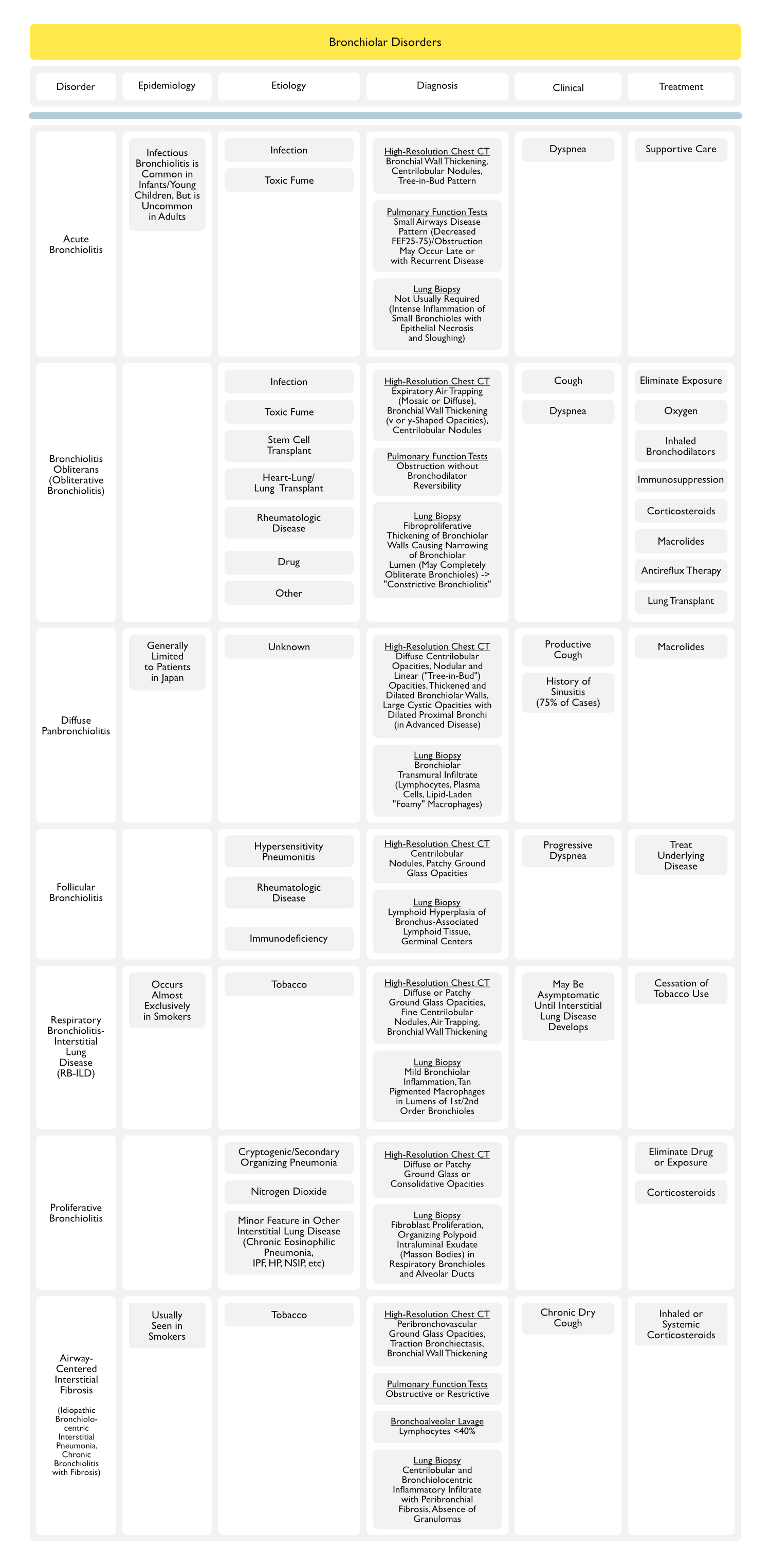
(aka Smoker’s Bronchiolitis)
Epidemiology
- Some cases have been reported in collagen vascular diseases and mineral dust exposure
- Time course: symptoms usually present for average of 37 months before presentation (typically, this is a longer course than for BOOP)
- Mean Age of Onset: 40’s
- Relationship to Smoking: almost all cases are smoking-related
Physiology
- Accumulation of pigmented alveolar macrophages in respiratory bronchioles and adjacent alveoli
- May represent a precursor to chronic lung disease in heavy smokers
Pathology
- Tan-brown pigmented macrophages (“smoker’s macrophages”) within respiratory bronchioles, alveolar ducts, and alveoli/ mildly thickened brinchiole wall with ectasia (with mucous stasis)/ extension of metaplastic bronchiolar epithelium into nearby alveoli
- Distant uninvolved parenchyma is normal or may demonstrate mild hyperinflation
- Initial cases reported were probably misclassified as DIP (due to DIP-like appearance)
Diagnosis
- ABG: mild hypoxemia
- PFT’s: usually mild-moderate restriction with normal-slightly decreased DLCO (normal PFT’s in some cases)
- Mixed obstructive/restrictive pattern: common
- Isolated increased RV: occasionally seen
- CXR/Chest CT Patterns
- Diffuse fine reticulonodular infiltrates: with normal lung volumes
- Bronchial wall thickening/ prominence of peribronchovascular interstitium/ small regular and irregular opacities/ small peripheral ring shadows are dustinctive features
Clinical
(insidious onset)
- Dyspnea:
- Cough (common):
- Rales: coarse, end-inspiratory
- Wheezing: reported in some cases
- Absence of clubbing
Treatment
- Steroids: favorable response in most series (improved PFT’s and CXR)
- Smoking cessation: leads to improvement in symptoms, PFT’s, and CXR
Prognosis
- No deaths have been reported
References
- xxx