In Emergency Endotracheal Intubations Performed in Critically Ill Patients Suffering Deterioration, the Rate of Cardiac Arrest was 1 in 50 and was Associated with Hypoxemia in 83% of Cases (and 63% of the Cases of Hypoxemia were Associated with Inadvertent Esophageal Intubation) (Anesth Analg, 2004) [MEDLINE]
In Emergency Department Intubations, Early Post-Intubation Cardiac Arrest Occurred in Approximately 2% of Cases and was Associated with Preintubation Systolic Hypotension (PLoS One, 2014) [MEDLINE]
In Intubations of Critically Ill Intensive Care Unit Patients, Post-Intubation Cardiac Arrest Occurred in 1 of 40 Procedures, Had High Immediate and 28-Day Mortality Rates, and was Associated with Defined Risk Factors (Crit Care Med, 2018) [MEDLINE]
Absence of Preoxygenation: odds ratio 3.584 (1.287-9.985)
Pre-Intubation Hypotension (SBP <90 mm Hg): odds ratio 3.406 (1.797-6.454)
Pre-Intubation Hypoxemia: odds ratio 3.991 (2.101-7.583)
Age > 75 y/o: odds ratio 2.251 (1.080-4.678)
Obesity (BMI>25): odds ratio 2.005 (1.017-3.951)
Reverse Shock Index Predicts Post-Intubation Cardiac Arrest (Int J Emerg Med, 2023) [MEDLINE]: n = 127
Reverse Shock Index = systolic blood pressure/heart rate
In Multivariate Analysis, Age, Reverse Shock Index, Oxygen Saturation, and Total Bilirubin were Significantly Associated with Post-Intubation Cardiac Arrest
Patients with Reverse Shock Index <1 had a Significantly Higher Risk of Developing Post-Intubation Cardiac Arrest (Odds Ratio 5.22; 95% CI: 1.83-14.86, p = 0.002)
Reverse Shock Index Sensitivity, Specificity, Positive Predictive Value, Negative Predictive Value, and Diagnostic Accuracy for Predicting Post-Intubation Cardiac Arrest were 51.11%, 83.33%, 90.2%, 36.23%, and 59.17%, Respectively
ROC Curve for Reverse Shock Index Showed an Area Under the Curve (AUC) of 0.66
Typically Occurs Due to Initiation of Positive-Pressure Ventilation in a Patient with Shock
Pulseless Electrical Activity was the Most Common Rhythm in Post-Intubation Early Cardiac Arrest (Occurred in 78.1% of Cases) (PLoS One, 2014) [MEDLINE]
Airway Obstruction with Expiratory Flow Limitation (Due to Asthma, COPD, etc) (Crit Care Med, 2000) [MEDLINE]
Expiratory Flow Limitation Impairs Exhalation and Lung Emptying
Expiratory Flow Limitation is More Prominent in the Supine Position than in the Semirecumbent Position (Am J Respir Crit Care Med, 1994) [MEDLINE]
Expiratory Flow Resistance (Due to Small Endotracheal Tube, Kinked Endotracheal Tube, Ventilator Tubing, Exhalation or PEEP Valve, or Patient-Ventilator Dyssynchrony)
Expiratory Resistance Impairs Exhalation and Lung Emptying
High Minute Ventilation (VE)
High Minute Ventilation (VE) May Due to Any/All of the Following
High Set Respiratory Rate (RR) on the Ventilator
High Patient-Driven Respiratory Rate (RR) (Due to Fever, Sepsis, Pain, Agitation, Anxiety, etc)
High Set Tidal Volume (VT) on the Ventilator
High Patient-Driven Tidal Volume (VT) (Due to Fever, Sepsis, Pain, Agitation, Anxiety, etc)
High Inspiratory/Expiratory (I/E) Ratio
While the “Normal” I/E Ratio for a Normal Spontaneously-Ventilating Patient is 1:2, a “High” I/E Ratio Cannot Be Numerically Defined for All Patients, Since Patients with Significant Airway Resistance (i.e. Airway Obstruction Due to Status Asthmaticus, etc) May Develop Auto-PEEP Even at a Relatively “Normal” I/E Ratio
Time-Constant Inequality of Lung Units
Some Lung Units (Especially in the Setting of Airway Obstruction) Empty Heterogeneously, Resulting in the Development of Auto-PEEP Even at a Relatively Low Minute Ventilation (VE)
Altered Respiratory System Compliance (Due to Expiratory Muscle Activity): impairs exhalation
Altered Respiratory System Compliance May Also Interfere with the Accurate Measurement of Auto-PEEP (Am J Respir Crit Care Med, 1995) [MEDLINE]
Physiology
Next Ventilator Breath is Triggered Before the Airway Pressure Returns to Baseline (i.e. Zero)
Unlike Applied PEEP Which Distributes Evenly, Auto-PEEP Distributes Predominantly to Lung Units with the Highest Airway Resistance and Lowest Compliance (Chest, 1995) [MEDLINE]
Diagnosis
Auscultation (or Palpation) of Continued Expiratory Airflow at the Point When the Next Ventilator Breath is Initiated
Physical Exam Has a Positive Predictive Value of 95% and a Negative Predictive Value of 58% in Detecting the Presence of Auto-PEEP, Suggesting that it is Useful to Diagnose Auto-PEEP, But Not to Rule Out Auto-PEEP (Am J Respir Crit Care Med, 1999) [MEDLINE]
Observation of Pressure Waveform Not Returning to Baseline Between Breaths
Expiratory Hold (0.5-1.0 sec) on Ventilator (i.e. Occlusion of Exhalation Port on Ventilator)
Clinical Effects
Dynamic Hyperinflation Amplifies the Respiratory Variation of Arterial Pulse Pressure and Contributes to Pulsus Paradoxus in Mechanically Ventilated Patients [MEDLINE]
Due to Decreased Venous Return to the Right Side of the Heart, Resulting in Decreased Cardiac Output (Especially in the Setting of Hypovolemia)
Increased Risk of Barotrauma (Mainly Due to Increased Lung Volume): see below
Increased Work of Breathing
Due to the Fact that the Patient Must Overcome the Residual Positive Airway Pressure to Generate a Negative Pressure Deflection to Trigger the Next Ventilator Breath
Due to Ventilation at High Lung/Chest Wall Volumes (Lung and Chest Wall are Less Compliant at High Lung Volumes)
Introduction of Errors in the Measurement of the Mean Alveolar Pressure and Static Lung Compliance (Am J Respir Crit Care Med, 1996) [MEDLINE]
Introduction of Errors in the Measurement of Pulmonary Capillary Wedge Pressure (PCWP) (see Hemodynamics)
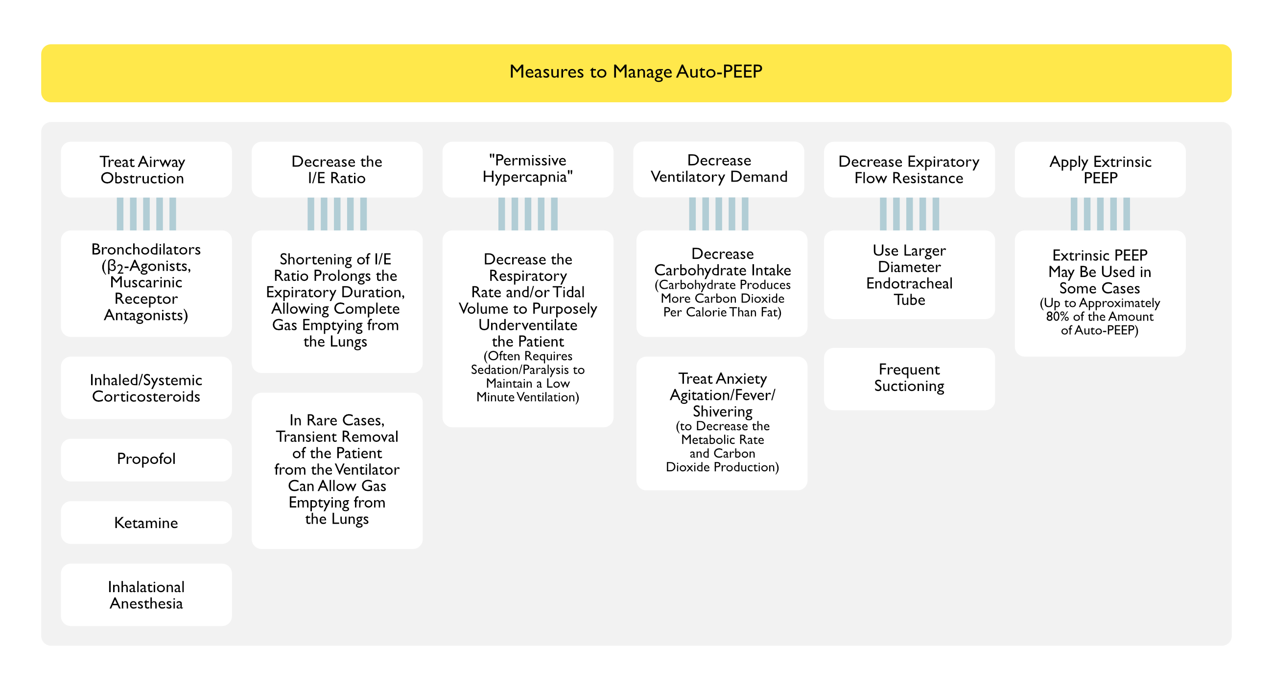
Increase the Inspiratory Flow Rate and/or Decrease the Respiratory Rate (RR) to Decrease the Inspiratory/Expiratory (I/E) Ratio (to 1:3, 1:4, 1:5, etc)
This Lengthens the Expiratory Time, Allowing a Longer Duration to Expire the Gas in the Lung
In Rare Cases, Transient Removal of the Patient from the Ventilator May Allow Gas Emptying from the Lungs
“Permissive Hypercapnia” (Decrease the Respiratory Rate and/or Tidal Volume to Purposely Underventilate the Patient): allowing the pCO2 to increase to as much as 70-100 mm Hg
Physiology
Permissive Hypercapnia was First Utilized in Status Asthmaticus
Permissive Hypercapnia Decreases the Tidal Volume (VT) (Which Will Decrease the Total Volume of Gas Which Must Be Exhaled During Expiration) and the Respiratory Rate (Which Will Allow a Longer Expiratory Time)
Maintenance of pH >7.2 Can Be Achieved with Either Sodium Bicarbonate (or Tris-Hydroxymethyl Aminomethane, THAM) Administration: however, sodium bicarbonate administration may be ineffective in increasing the pH in this setting
Many Patients Require Deep Sedation (and Paralysis, if Necessary) to Maintain a Low Minute Ventilation (VE)
Measures to Decrease the Metabolic Rate (Decreasing Carbon Dioxide Production and Therefore, the Ventilatory Demand)
In Patients with Underlying Reactive Airways Disease, Postoperative Bronchospasm Occurs Commonly with Endotracheal Intubation (Masui, 1995) [MEDLINE]: bronchospasm occurs in 8.9% of patients with reactive airways disease undergoing endotracheal intubation with general anesthesia
In Patients with Underlying Reactive Airways Disease, the Incidence of Postoperative Bronchospasm was Higher with Thoracic/Abdominal Surgery (39.5%), as Compared to Other Surgeries (10.4%) (Masui, 1995) [MEDLINE]
Mechanisms
Aspiration of Acidic Gastric Contents During Intubation, Resulting in Airway Irritation
Check Endotracheal Tube and Ventilator Circuit for Disconnections or Leaks
Monitor Endotracheal Tube Cuff Pressure with Manometer
Bronchoscopy (see Bronchoscopy): useful to rapidly determine the location of the endotracheal tube tip, in cases where endotracheal tube proximal malpositioning is suspected
With a Suspected Cuff Leak, if the Endotracheal Tube Tip is in the Appropriate Position, This Makes the Diagnosis of Endotracheal Tube Cuff Rupture More Likely as the Etiology
Clinical
Endotracheal Tube Cuff Leak
In One Study of Patients with Lost Volume on the Ventilator and Suspected Endotracheal Tube Cuff Rupture, 61% of the Leaks Were Actually Due to Endotracheal Tube Dislodgment and Only 39% Were Due to a Ruptured Cuff (Crit Care Med, 1993) [MEDLINE]
Audible Leak from Air Passing Through Around the Cuff of the Endotracheal Tube
Decreased Peak Airway Pressure (PIP) on Ventilator
Extent and Leak and Patient Tolerance of the Endotracheal Tube Cuff Leak are Variable (Anesth Analg, 2013) [MEDLINE]
Inability to Maintain Endotracheal Tube Cuff Pressure
“Lost Volume” on the Ventilator (Inspired Tidal Volume > Expired Tidal Volume)
Ventilator Circuit Leak
Decreased Peak Airway Pressure (PIP) on Ventilator
“Lost Volume” on the Ventilator (Inspired Tidal Volume > Expired Tidal Volume)
Treatment
Endotracheal Tube Cuff Leak Due to a Ruptured/Damage Endotracheal Tube Cuff
Requires Reintubation
Endotracheal Tube Cuff Leak Due to Damaged Pilot Ballon Tubing
Pilot Ballon Tubing Can Be Repaired by Cutting the Pilot Ballon Line Prior to Site of Leak (If Feasible) and Repaired Using a Commercially-Available Kit
Leak in Pilot Ballon Tubing Can Also Be Repaired by Cutting the Pilot Ballon Line Prior to Site of Leak (If Feasible) and Using a Peripheral Intravenous Catheter (BMC Anesthesiol, 2022) [MEDLINE]
Time to Repair Leak: 27.8 ± 1.5 s in the endotracheal tube group (and 20.4 ± 1.1 s in the laryngeal mask airway group)
Reintubation is Required in Cases Where the Leak Cannot Be Remedied Using One of the Above Methods
Ventilator Circuit Leak
Replace Ventilator Circuit Tubing
Endotracheal Tube Tip Malpositioning
Epidemiology
In One Study of Patients with Lost Volume on the Ventilator and Suspected Endotracheal Tube Cuff Rupture, 61% of the Leaks Were Actually Due to Endotracheal Tube Dislodgment and Only 39% Were Due to a Ruptured Cuff (Crit Care Med, 1993) [MEDLINE]
Mechanisms
Inadvertent Placement of the Endotracheal Tip Either Proximally (i.e. Endotracheal Tube Cuff Above the Vocal Cords) or Distally (i.e. Mainstem Bronchial Intubation)
Diagnosis
Bronchoscopy (see Bronchoscopy): useful to rapidly identify the endotracheal tube tip location
Thoracic Ultrasound (see Thoracic Ultrasound): useful to confirm endotracheal tube position
Requirement for Increasing Volume to Maintain Endotracheal Tube Cuff Pressure: caution should be exercised in cases such as these, as unrecognized excessive inflation of the cuff may occur in the posterior oropharynx in a patient with a proximally-migrated endotracheal tube
Unilaterally Absent Breath Sounds (on the Non-Ventilated Side)
Pneumothorax (on the Overventilated Side) (see Pneumothorax): use of large tidal volume delivered unilaterally may particularly predispose to the development of pneumothorax on the ventilated side
Pneumothorax (see Pneumothorax): use of large tidal volume delivered unilaterally may particularly predispose to the development of pneumothorax on the ventilated side
Kinked Endotracheal Tube
Mechanism
Kinking at Endotracheal Tube Securement Device (Hollister, etc): common
Kinking in Posterior Oropharynx: less common
Kinking at Teeth (i.e. Patient Biting the Endotracheal Tube): most common site of kinking
Diagnosis
Bronchoscopy (see Bronchoscopy): useful to rapidly evaluate endotracheal tube patency
Clinical
Increased Peak Airway Pressure (PIP) on Ventilator
Post-Intubation Hypotension is the Most Common Complication of Intubation
Up to 40% of Patients Intubated in the ICU Setting Experience Significant Procedure-Related Hypoxemia or Hypotension (Crit Care, 2015) [MEDLINE]
Risk Factors for Post-Intubation Hypotension (Crit Care, 2015) [MEDLINE]
Simplified Acute Physiologic Score II (SAPS II): odds ratio 1.02 (p<0.001)
Age 60-75 y/o: odds ratio 1.96 (p<0.002 vs <60 y/o)
Age >75 y/o: odds ratio 2.81 (p<0.001 vs <60 y/o)
Acute Respiratory Failure as the Indication for Intubation: odds ratio 1.51 (p=0.04)
First Intubation in the ICU: odds ratio 1.61 (p=0.02)
Noninvasive Ventilation Required for Preoxygenation: odds ratio 1.54 (p=0.03)
Inspired FIO2 >70% After Intubation: odds ratio 1.91 (p=0.001)
Mechanisms
Positive-Pressure Ventilation Increases Intrathoracic and Right Atrial Pressure -> Decreases Venous Return to the Right Side of the Heart -> Decreases Right Ventricular Cardiac Output
This Effect is Accentuated by the Concomitant Presence of Auto-PEEP, Extrinsic PEEP, and/or Hypovolemia (Anesthesiology, 1975) [MEDLINE]
The Use of Pharmacologic Agents with Vasodilator Properties (Opiates, Benzodiazepines, Propofol, etc) Can Further Exacerbate this Effect
This Effect May Be Most Pronounced Immediately After Intubation and Initiation of Mechanical Ventilation in a Patient Who is Hypovolemic and Has Just Received Vasodilating Sedatives (Such as Midazolam, Propofol, etc) or Analgesics (Fentanyl, etc)
Positive-Pressure Ventilation Causes Alveolar Inflation with Compression of the Pulmonary Vascular Bed -> Increases Pulmonary Vascular Resistance (PVR) -> Decreases Right Ventricular Output (Crit Care Med, 2010) [MEDLINE]
Passive Leg Raise Maneuver Has Been Demonstrated to Increase Central Blood Volume and Mitigate this Effect (Crit Care Med, 2010) [MEDLINE]
Positive-Pressure Ventilation Causes Alveolar Inflation with Compression of the Pulmonary Vascular Bed -> Increases Pulmonary Vascular Resistance (PVR) -> Shifts the Intraventricular Septum Toward the Left (with Impaired Diastolic Left Ventricular Filling) -> Decreases Left Ventricular Cardiac Output
Peri-Intubation Pneumothorax (see Pneumothorax): may occur due to large tidal volume during bag-valve-mask ventilation
Prophylactic Intravenous Fluid/Vasopressors Prior to and/or During Endotracheal Intubation
Especially Indicated in Patients with Marginal Pre-Intubation Blood Pressure and/or Known Hypovolemia
PREPARE II Randomized Trial of Intravenous Fluid Bolus Prior to Endotracheal Intubation (JAMA, 2022) [MEDLINE]: n = 1,067 (in 11 intensive care units in the United States)
In Critically Ill Adults Undergoing Endotracheal Intubation, Administration of an Intravenous Fluid Bolus (as Compared to No Fluid Bolus) Did Not Significantly Decrease the Incidence of Cardiovascular Collapse or 28-Day Mortality Rate
In an Analysis of Data from the Prospective, Multicenter, Observational Japanese Emergency Airway Network (JEAN-2) Study (from Feb, 2012-Nov 2017), Ketamine Manifested Less Post-Intubation Hypotension in Hemodynamically-Unstable Patients in the Emergency Department, as Compared to Midazolam or Propofol (Sci Rep, 2019) [MEDLINE]
Paralysis with Inadequate Sedation: this may especially occur when a short-acting sedative (such as etomidate, etc) is utilized in conjunction with a long-acting paralytic (such as rocuronium, etc)
Positive-Pressure Ventilation in Hypovolemic Patient, Resulting in Decreased Venous Return to Right Side of the Heart with Decreased Cardiac Output
Sympathetic Nervous System Response Due to Glottic Stimulation: see below
Sympathetic Nervous System Response
Mechanisms
Glottic Stimulation from Laryngoscopy Blade/Endotracheal Tube (Typically in the Setting of Inadequate Sedation): since the glottis is highly innervated
Paralysis with Inadequate Sedation: may occur following rapid sequence intubation with a short-acting sedative (such as etomidate, etc) and a long-acting paralytic (such as rocuronium, etc)
Note that Sinus Tachycardia May Alternatively Represent a Normal Physiologic Response to Hypovolemia Induced by Positive-Pressure Ventilation (and Decreased Venous Return to the Right Side of the Heart): it is critical to rule out hypovolemia as the etiology in this case
Prevention of Sympathetic Nervous Response Due to Glottic Stimulation
Bronchoscopy is Recommended to Rapidly Determine the Site of Post-Intubation Hemoptysis (Larynx vs Tracheal Mucosal vs Endobronchial Mucosal vs Diffuse Alveolar Hemorrhage): this is especially useful in patients with pre-existing diffuse alveolar hemorrhage who only manifest hemoptysis after intubation
Ventilator-Induced Lung Injury (VILI): lung injury due to volume-related overstretching (“volutrauma”), high frequency of stretching, and/or high velocity/acceleration of stretching
VILI Likely Develops Regionally in the Lung When Low Resistance/High Compliance Lung Units Receive a Disproportionately Large Regional Tidal Volume in the Setting of High Alveolar Distending Pressures
Pathologically, VILI Appears as Diffuse Alveolar Damage
VILI is Associated with Cytokine Release and Bacterial Translocation
Barotrauma: clinically apparent type of alveolar injury which presents as extra-alveolar air in various locations (mediastinum, pleural space, etc)
Epidemiology
Mechanical Ventilation Itself Increases the Risk of Barotrauma
Development of Auto-PEEP During Mechanical Ventilation Further Increases the Risk of Barotrauma
Noninvasive Positive-Pressure Ventilation Probably Has a Similar Mechanism of Barotrauma as Invasive Mechanical Ventilation, But the Rate of Barotrauma is Lower (Due to Use of Lower Pressures) (Rev Bras Ter Intensiva, 2008) [MEDLINE]
Incidence of Barotrauma in ARDS is Approximately 10% (NEJM, 2000) [MEDLINE] (Intensive Care Med, 2002) [MEDLINE] (NEJM, 2004) [MEDLINE]
Physiology
Ventilator Factors Which May Cause Alveolar Overdistention, Resulting in Alveolar Rupture
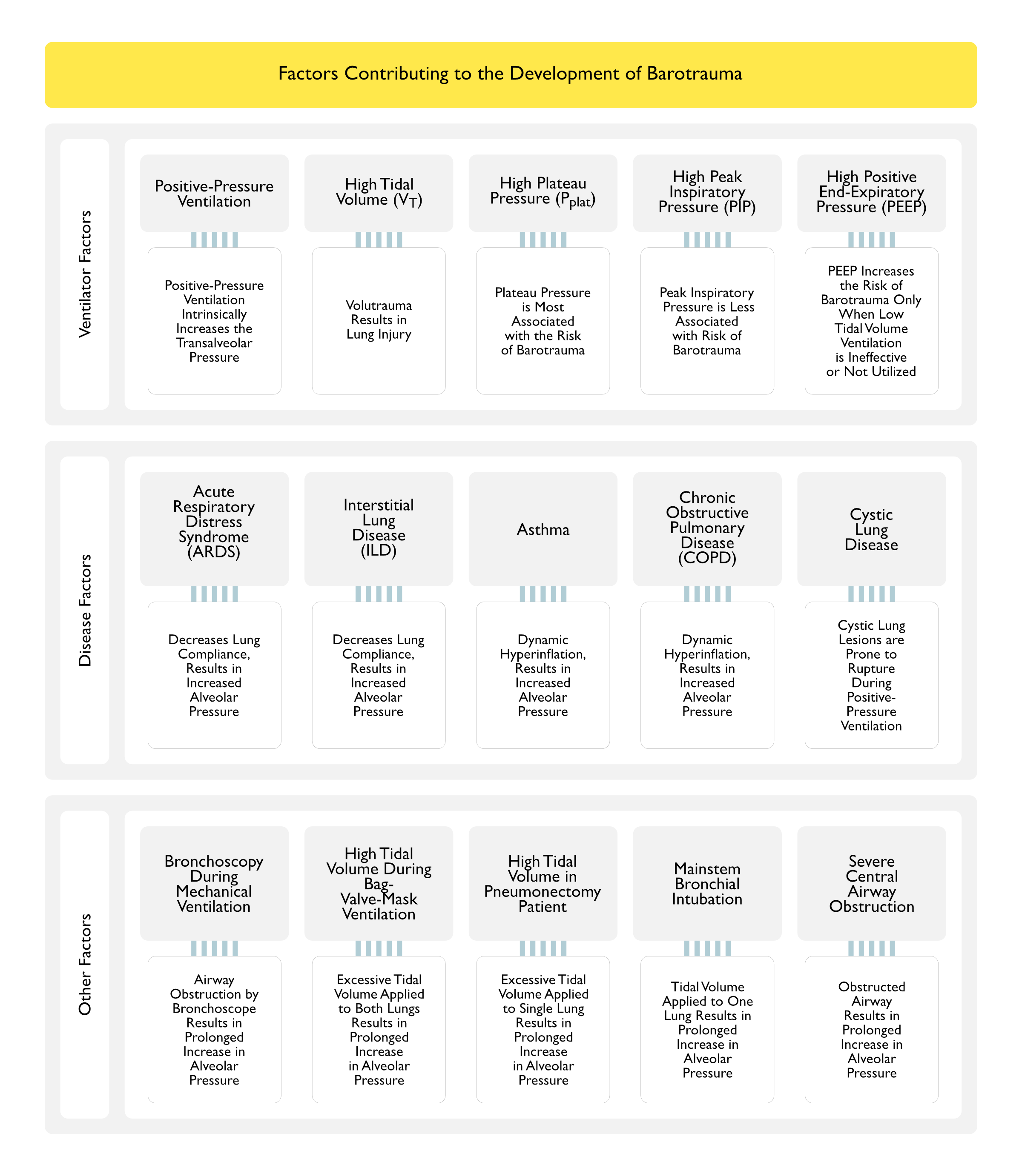
Positive-Pressure Ventilation Itself
All Patients on Mechanical Ventilation are at Risk for Barotrauma, Since Positive-Pressure Ventilation Increases the Transalveolar Pressure (Alveolar Pressure – Adjacent Interstitial Space Pressure)
High Tidal Volume
Inappropriately High Tidal Volume Set During Bag-Valve-Mask Ventilation
Inappropriately High Tidal Volume Set on Ventilator (During Volume-Cycled Ventilation)
Inadvertent Right (or Left) Mainstem Intubation with Inappropriately High Tidal Volume Applied to a Single Lung
High Plateau Pressure (Pplat)
Plateau Pressure is the Pressure Applied to the Small Airways and Alveoli
While There is No Safe Plateau Pressure Under Which Barotrauma Does Not Occur, the Greatest Risk of Barotrauma Occurs with Plateau Pressure ≥35 cm H2O or Static Compliance <30 mL/cm H2O (Intensive Care Med, 2002) [MEDLINE]
High Peak Inspiratory Pressure (PIP)
There is No Absolute Safe Threshold for PIP Under Which Barotrauma Does Not Occur
PIP is Probably Less Associated with the Risk of Barotrauma than the Plateau Pressure, Although Some Data are Conflicting (Crit Care Med, 1983) [MEDLINE] (Chest, 1994) [MEDLINE] (Am J Respir Crit Care Med, 2002) [MEDLINE]
High Positive End-Expiratory Pressure (PEEP)
High PEEP Probably Only Contributes to an Increased Risk of Barotrauma When Open Lung Ventilation (High PEEP or Recruitment Maneuvers, Usually with Lung Protective Measure Such as Low Tidal Volume with Plateau Pressure ≤30 cm H2O) Strategies are Ineffective in Recruiting Atelectatic Lung Units in ARDS or When Lung Protective Ventilation (Low Tidal Volume with Plateau Pressure ≤30 cm H2O) is Not Utilized
Mode of Ventilation (Volume-Cycled vs Pressure-Cycled) Does Not Appear to Be Associated with the Risk of Barotrauma (PLoS One, 2011) [MEDLINE] (Cochrane Database Syst Rev, 2015) [MEDLINE]
Disease Factors Which May Cause Alveolar Overdistention, Resulting in Alveolar Rupture
Acute Respiratory Distress Syndrome (ARDS): due to decreased lung compliance, resulting in increased alveolar pressure (Am J Respir Crit Care Med, 1995) [MEDLINE] (Intensive Care Med, 2004) [MEDLINE]
Asthma (see Asthma): due to dynamic hyperinflation, resulting in increased alveolar pressure (Intensive Care Med, 2004) [MEDLINE]
Chronic Obstructive Pulmonary Disease (COPD) (see Chronic Obstructive Pulmonary Disease): due to dynamic hyperinflation, resulting in increased alveolar pressure
Interstitial Lung Disease (ILD) (see Interstitial Lung Disease): due to decreased lung compliance, resulting in increased alveolar pressure (Intensive Care Med, 2004) [MEDLINE]
Langerhans Cell Histiocytosis (see Langerhans Cell Histiocytosis): due to cystic lung disease, resulting in escape of air
Pneumocystis Jirovecii Pneumonia (see Pneumocystis Jirovecii): due to cavitating lung disease, resulting in escape of air
Other Factors Which May Cause Alveolar Overdistention, Resulting in Alveolar Rupture
Bronchoscopy During Mechanical Ventilation: may result in prolonged increase in plateau pressure
High Tidal Volume During Bag-Valve-Mask Ventilation: may result in prolonged increase in plateau pressure
High Tidal Volume Ventilation in Pneumonectomy Patient: may result in prolonged increase in plateau pressure
Right Mainstem Bronchial Intubation: may result in prolonged increase in plateau pressure
Severe Central Airway Obstruction: may result in prolonged increase in plateau pressure
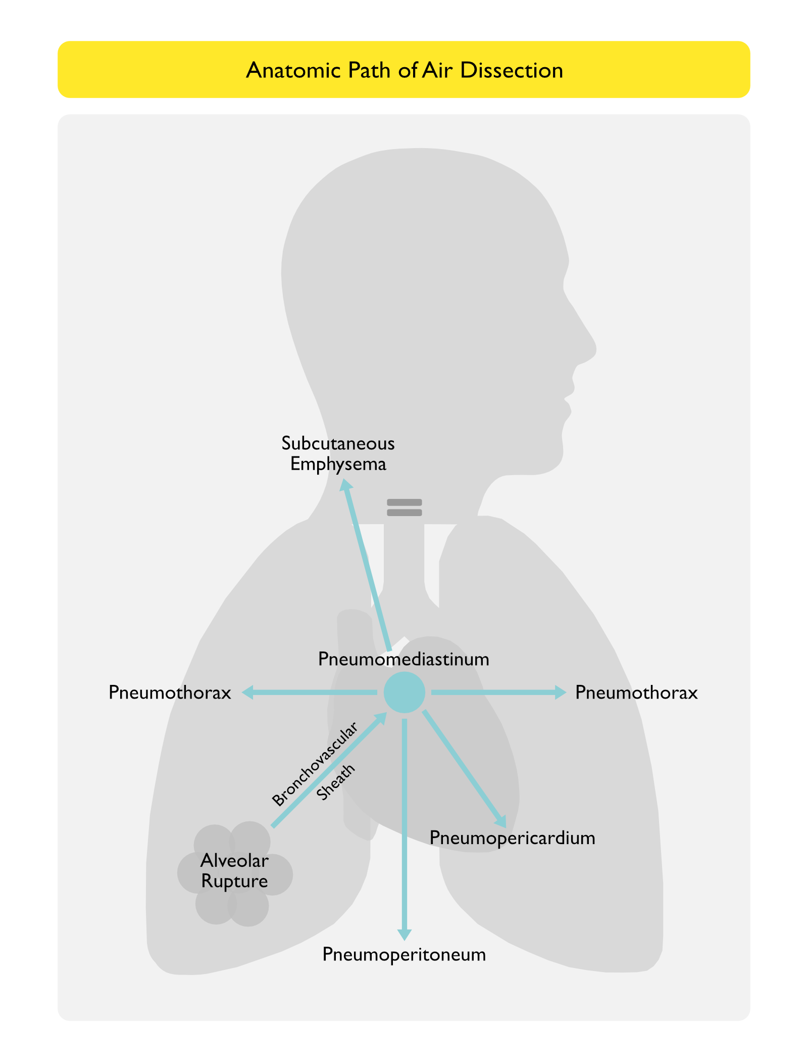
Anatomic Path of Air Dissection
Air from Torn Alveolus Enters the Perivascular Interstitium, Dissecting Along the Bronchovascular Sheath into the Pulmonary Hila and Subsequently Into the Mediastinum, Causing Pneumomediastinum (in the Setting of Blunt Trauma to the Lung, This Tracking of Air Has Been Termed the “Macklin Effect”) (see Pneumomediastinum) (Chest, 2001) [MEDLINE]
From Pneumomediastinum, Air Can Dissect Upward into the Soft Tissues of the Neck (Causing Subcutaneous Emphysema), into the Pleural Spaces (Causing Pneumothorax on Either Side), Inferiorly into the Peritoneum (Causing Pneumoperitoneum), or Rarely, into the Pericardium (Causing Pneumopericardium)
Diagnosis
Bronchoscopy (see Bronchoscopy): useful to rapidly identify and treat large airway mucous plugging and identify endotracheal tube tip location
While Both Barotrauma and Volutrauma Likely Contribute to Alveolar Injury, Limiting Alveolar Pressure Appears to Be the Most Effective Measure to Prevent Barotrauma
Avoid Dynamic Hyperinflation (in Asthma and COPD): hyperinflation is progressive (dynamic) since air accumulates in the lung with each breath as a result of a failure to achieve complete exhalation before the onset of the next breath
Treatment of Bronchospasm (see Obstructive Lung Disease): usually with bronchodilators, corticosteroids, etc
Use Low Tidal Volume Ventilation: reduces amount of air in each breath which needs to be exhaled
Use Short Inspiratory Time/Longer Expiratory Time: allows adequate time for expiration
Use Lower Respiratory Rate (Even Using Permissive Hypercapnia in Some Cases)
Maintain Low Plateau Pressure (Pplat ≤30 cm H2O): being cautious to avoid Pplat >35 cm H2O
Targeting Even Lower Plateau Pressures May Further Reduce the Risk of Barotrauma (Am J Respir Crit Care Med, 1999) [MEDLINE]
In in Systematic Review and Meta-Analysis, Use of Neuromuscular Blockade is Associated with a Decreased Risk of Barotrauma (Crit Care, 2013) [MEDLINE]
Use Appropriate Amounts of PEEP
PEEP is Standardly Titrated Per the FIO2/PEEP Table as was Used in the 2000 Study on Low Tidal Volume Ventilation (N Engl J Med, 2000) [MEDLINE]
Use Low Respiratory Rate: even using permissive hypercapnia in some cases
Management-General
Since the Occurrence of Any Barotrauma in the Setting of Mechanical Ventilation for ARDS Suggests Either a Patient with More Severe ARDS (at Higher Mortality Risk) or a Suboptimal Ventilation Strategy (or Both), the First Priority Should Be Measures to Optimize the Ventilation Management
Use Lung Protective/Low Tidal Volume Ventilation (Ideally to 6 mL/kg PBW) and Adjust the Respiratory Rate to the Minimum Required to Maintain an Adequate pH (i.e. Permissive Hypercapnia)
Use Sedation and Pharmacologic Paralysis as Necessary to Maintain Ventilator Parameters
Avoid Patient-Ventilator Dyssynchrony and Overbreathing
Use Sedation and Pharmacologic Paralysis as Necessary to Maintain Synchrony
Decrease Both the Auto-PEEP and Extrinsic PEEP (As Allowed by Oxygenation)
Target a Low I/E Ratio (Around 0.33) to Maintain a Low Mean Airway Pressure
Shorten Inspiratory Time with Higher Inspiratory Flow Rate (Around 70-100 L/min)
Lengthen Expiratory Time
Avoid Inverse Ratio Ventilation
Use a Low Compressible Volume (Non-Disposable) Ventilator Circuit
Chest Tube is Generally Required for Mechanical Ventilation-Associated Pneumothorax, Since >30% of These Progress to Tension Pneumothorax
Management-Chest Tube with Air Leak (i.e. Bronchopleural/Alveolopleural Fistula) (see Bronchopleural Fistula): ventilator changes should aim to decrease the plateau pressure (to Pplat ≤30 cm H2O)
Optimization of Ventilation Management: as above
Continue Chest Tube Drainage (see Chest Tube: while this may seem obvious, prematurely removing a chest tube in a patient with an air leak from a bronchopleural fistula can result in the rapid development of tension pneumothorax (which can be potentially fatal)
Use the Least Amount of Chest Tube Suction Which Maintains Lung Inflation and Decreases the Amount of Air Leak
Optimize Body Position and Patient’s Sedation/Pharmacologic Paralysis to Minimize the Air Leak
Barotrauma-Associated Pneumoperitoneum is Usually Self-Limited and Doesn’t Require Specific Intervention (Other than Ventilator Adjustments, Such as a Decrease in Plateau Pressure, etc)
Rare Cases of Pneumoperitoneum-Associated Abdominal Compartment Syndrome May Require Lapartotomy/Laparoscopy for Surgical Decompression (to Both Relieve the Compartment Syndrome and to Exclude a Perforated Viscus)
In Mixed Populations, Mechanical Ventilation-Associated Barotrauma is Associated with an Increased Mortality Rate (Chest, 1986) [MEDLINE]
ARDS-Associated Barotrauma is Associated with an Increased Mortality Rate: likely related to the fact that the barotrauma is a marker for patients with worse ARDS (Crit Care Med, 1995) [MEDLINE] (JAMA, 1994) [MEDLINE]
BEST Kidney Trial. Acute renal failure in critically ill patients: a multinational, multicenter study. JAMA. 2005;294(7):813 [MEDLINE]
Mechanical ventilation and acute renal failure. Crit Care Med. 2005;33(6):1408 [MEDLINE]
Arrhythmia/Cardiac Arrest (see Cardiac Arrest)
Emergency tracheal intubation: complications associated with repeated laryngoscopic attempts. Anesth Analg. 2004 Aug;99(2):607-13, table of contents [MEDLINE]
Factors associated with the occurrence of cardiac arrest after emergency tracheal intubation in the emergency department. PLoS One. 2014 Nov 17;9(11):e112779. doi: 10.1371/journal.pone.0112779. eCollection 2014 [MEDLINE]
Cardiac Arrest and Mortality Related to Intubation Procedure in Critically Ill Adult Patients: A Multicenter Cohort Study. Crit Care Med. 2018 Apr;46(4):532-539. doi: 10.1097/CCM.0000000000002925 [MEDLINE]
Shock Index as a Predictor of Post-Intubation Hypotension and Cardiac Arrest; A Review of the Current Evidence. Bull Emerg Trauma. 2019 Jan;7(1):21-27. doi: 10.29252/beat-070103 [MEDLINE]
Reverse shock index (RSI) as a predictor of post-intubation cardiac arrest (PICA). Int J Emerg Med. 2023 Dec 7;16(1):88. doi: 10.1186/s12245-023-00569-y [MEDLINE]
Arytenoid Cartilage Dislocation
Cardiovascular operation: A significant risk factor of arytenoid cartilage dislocation/subluxation after anesthesia. Ann Card Anaesth. 2017 Jul-Sep;20(3):309-312. doi: 10.4103/aca.ACA7117 [MEDLINE]
Arytenoid cartilage dislocation mimicking bilateral vocal cord paralysis: A case report. Medicine (Baltimore). 2017 Nov;96(45):e8514. doi: 10.1097/MD.0000000000008514 [MEDLINE]
Unusual cause of hoarseness: Arytenoid cartilage dislocation without a traumatic event. Am J Emerg Med. 2018 Jan;36(1):172.e1-172.e2. doi: 10.1016/j.ajem.2017.10.041 [MEDLINE]
BMI May Be the Risk Factor for Arytenoid Dislocation Caused by Endotracheal Intubation: A Retrospective Case-Control Study. J Voice. 2018 Mar;32(2):221-225. doi: 10.1016/j.jvoice.2017.05.010 [MEDLINE]
Aspiration Pneumonia (see Aspiration Pneumonia)
Swallowing disorders in patients with prolonged orotracheal intubation or tracheostomy tubes. Crit Care Med. 1990 Dec;18(12):1328-30. doi: 10.1097/00003246-199012000-00004 [MEDLINE]
Effect of positive expiratory pressure and type of tracheal cuff on the incidence of aspiration in mechanically ventilated patients in an intensive care unit. Crit Care Med. 2008;36(2):409 [MEDLINE]
CDC/NHSN surveillance definition of health care-associated infection and criteria for specific types of infections in the acute care setting. Am J Infect Control. 2008;36(5):309 [MEDLINE]
Occult positive end-expiratory pressure in mechanically ventilated patients with airflow obstruction: the auto-PEEP effect. Am Rev Respir Dis. 1982;126(1):166 [MEDLINE]
PEEP, auto-PEEP, and waterfalls. Chest. 1989 Sep;96(3):449-51 [MEDLINE]
Detection of expiratory flow limitation during mechanical ventilation. Am J Respir Crit Care Med. 1994;150(5 Pt 1):1311 [MEDLINE]
Expiratory muscle activity increases intrinsic positive end-expiratory pressure independently of dynamic hyperinflation in mechanically ventilated patients. Am J Respir Crit Care Med. 1995;151(2 Pt 1):562 [MEDLINE]
Mean airway pressure as an index of mean alveolar pressure. Am J Respir Crit Care Med. 1996;153(6 Pt 1):1825 [MEDLINE]
Clinical examination reliably detects intrinsic positive end-expiratory pressure in critically ill, mechanically ventilated patients. Am J Respir Crit Care Med. 1999;159(1):290 [MEDLINE]
Intrinsic positive end-expiratory pressure in mechanically ventilated patients with and without tidal expiratory flow limitation. Crit Care Med. 2000;28(12):3837 [MEDLINE]
Dynamic hyperinflation and auto-positive end-expiratory pressure: lessons learned over 30 years. Am J Respir Crit Care Med. 2011;184:756–762 [MEDLINE]
Association between aspirin use and deep venous thrombosis in mechanically ventilated ICU patients. J Thromb Thrombolysis. 2017 Oct;44(3):330-334. doi: 10.1007/s11239-017-1525-x [MEDLINE]
Efficacy of using an intravenous catheter to repair damaged expansion lines of endotracheal tubes and laryngeal masks. BMC Anesthesiol. 2022 Jul 26;22(1):238. doi: 10.1186/s12871-022-01776-5 [MEDLINE]
Endotracheal Tube Tip Malpositioning
Massive airway leaks: an analysis of the role of endotracheal tubes. Crit Care Med. 1993 Apr;21(4):518-21 [MEDLINE]
The effects of positive end-expiratory pressure on the splanchnic circulation. Intensive Care Med. 2000 Apr;26(4):361-3. doi: 10.1007/s001340051168 [MEDLINE]
GI complications in patients receiving mechanical ventilation. Chest. 2001 Apr;119(4):1222-41. doi: 10.1378/chest.119.4.1222 [MEDLINE]
Ileus (and Gastrointestinal Hypomotility) (see Ileus)
Gastroduodenal motility in mechanically ventilated critically ill patients: a manometric study. Crit Care Med. 1994 Mar;22(3):441-7. doi: 10.1097/00003246-199403000-00014 [MEDLINE]
GI complications in patients receiving mechanical ventilation. Chest. 2001 Apr;119(4):1222-41. doi: 10.1378/chest.119.4.1222 [MEDLINE]
Gastroduodenal motility in mechanically ventilated critically ill patients: a manometric study. Crit Care Med. 1994;22(3):441 [MEDLINE]
The effects of positive end-expiratory pressure on the splanchnic circulation. Intensive Care Med. 2000;26(4):361 [MEDLINE]
Effect of positive end-expiratory pressure on splanchnic perfusion in acute lung injury. Intensive Care Med. 2000;26(4):376 [MEDLINE]
GI complications in patients receiving mechanical ventilation. Chest. 2001;119(4):1222 [MEDLINE]
Impaired Mucociliary Motility
Mucociliary transport in ICU patients. Chest. 1994;105(1):237 [MEDLINE]
Effect of On-Demand vs Routine Nebulization of Acetylcysteine With Salbutamol on Ventilator-Free Days in Intensive Care Unit Patients Receiving Invasive Ventilation: A Randomized Clinical Trial. JAMA. 2018;319(10):993 [MEDLINE]
Mechanical ventilation triggers hippocampal apoptosis by vagal and dopaminergic pathways. Am J Respir Crit Care Med. 2013 Sep;188(6):693-702 [MEDLINE]
Induction of Inflammatory Response
Effect of mechanical ventilation on inflammatory mediators in patients with acute respiratory distress syndrome: a randomized controlled trial. JAMA. 1999;282(1):54 [MEDLINE]
Insulin Resistance
Physical inactivity rapidly induces insulin resistance and microvascular dysfunction in healthy volunteers. Arterioscler Thromb Vasc Biol. 2007;27(12):2650 [MEDLINE]
Early physical and occupational therapy in mechanically ventilated, critically ill patients: a randomised controlled trial. Lancet. 2009;373(9678):1874 [MEDLINE]
Joint Contracture
Joint contracture following prolonged stay in the intensive care unit. CMAJ. 2008;178(6):691 [MEDLINE]
Laryngeal Injury
Intubation lesions of the larynx. Br J Anaesth. 1978;50(6):587 [MEDLINE]
Postoperative sore throat: influence of tracheal tube lubrication versus cuff design. Can Anaesth Soc J. 1980;27(2):156 [MEDLINE]
Laryngeal injuries secondary to nasogastric tubes. Ann Otol Rhinol Laryngol. 1981;90(5 Pt 1):469 [MEDLINE]
The influence of endotracheal tube cuff design and cuff lubrication on postoperative sore throat. Anesthesiology. 1983;58(4):376 [MEDLINE]
Laryngotracheal injury due to endotracheal intubation: incidence, evolution, and predisposing factors. A prospective long-term study. Crit Care Med. 1983;11(5):362 [MEDLINE]
True vocal cord paralysis following intubation. Laryngoscope. 1985;95(11):1352 [MEDLINE]
Correlation of endotracheal tube size with sore throat and hoarseness following general anesthesia. Anesthesiology. 1987 Sep;67(3):419-21 [MEDLINE]
Laryngeal complications of prolonged intubation. Chest. 1989;96(4):877 [MEDLINE]
Evaluation of risk factors for laryngeal edema after tracheal extubation in adults and its prevention by dexamethasone. A placebo-controlled, double-blind, multicenter study. Anesthesiology. 1992;77(2):245 [MEDLINE]
Adverse respiratory events infrequently leading to malpractice suits. A closed claims analysis. Anesthesiology. 1991;75(6):932 [MEDLINE]
Evaluation of risk factors for laryngeal edema after tracheal extubation in adults and its prevention by dexamethasone: a placebo-controlled, double-blind, multicenter study. Anesthesiology 1992;77:245–251 [MEDLINE]
Surgical considerations in tracheal stenosis. Laryngoscope. 1992 Mar;102(3):237-43 [MEDLINE]
Resolution of laryngeal injury following translaryngeal intubation. Am Rev Respir Dis. 1992;145(2 Pt 1):361 [MEDLINE]
Airway considerations in the management of patients requiring long-term endotracheal intubation. Anesth Analg. 1992;74(2):276 [MEDLINE]
Massive airway leaks: an analysis of the role of endotracheal tubes. Crit Care Med. 1993;21(4):518 [MEDLINE]
Risk factors associated with prolonged intubation and laryngeal injury. Otolaryngol Head Neck Surg. 1994 Oct;111(4):453-9 [MEDLINE]
Mediastinitis and sepsis syndrome following intubation. Anaesthesia. 1994;49(10):883 [MEDLINE]
Association between reduced cuff leak volume and postextubation stridor. Chest. 1996;110(4):1035 [MEDLINE]
Gastroesophageal reflux in patients with subglottic stenosis. Arch Otolaryngol Head Neck Surg. 1998 May;124(5):551-5 [MEDLINE]
Airway injury during anesthesia: a closed claims analysis. Anesthesiology. 1999;91(6):170 [MEDLINE]
The cuff leak test to predict failure of tracheal extubation for laryngeal edema. Intensive Care Med. 2002;28(9):1267 [MEDLINE]
Post-extubation stridor in intensive care unit patients. Risk factors evaluation and importance of the cuff-leak test. Intensive Care Med. 2003;29(1):69 [MEDLINE]
How to identify patients with no risk for postextubation stridor? J Crit Care. 2004;19(1):23 [MEDLINE]
The endotracheal tube cuff-leak test as a predictor for postextubation stridor. Respir Care. 2005;50(12):1632 [MEDLINE]
Association of airway abnormalities and risk factors in 37 subglottic stenosis patients. Otolaryngol Head Neck Surg. 2006 Sep;135(3):434-7 [MEDLINE]
Intravenous injection of methylprednisolone reduces the incidence of postextubation stridor in intensive care unit patients. Crit Care Med. 2006;34(5):1345 [MEDLINE]
Laryngeal ultrasound: a useful method in predicting post-extubation stridor. A pilot study. Eur Respir J. 2006;27(2):384 [MEDLINE]
Dexamethasone to prevent postextubation airway obstruction in adults: a prospective, randomized, double-blind, placebo-controlled study. Crit Care. 2007;11(4):R72 [MEDLINE]
12-h pretreatment with methylprednisolone versus placebo for prevention of postextubation laryngeal oedema: a randomised double-blind trial. Lancet. 2007;369(9567):1083 [MEDLINE]
Age and comorbidity as risk factors for vocal cord paralysis associated with tracheal intubation. Br J Anaesth. 2007 Apr;98(4):524-30. Epub 2007 Mar 6 [MEDLINE]
Short-term effects of endotracheal intubation on voice. J Voice. 2007 Nov;21(6):762-8. Epub 2006 Aug 14 [MEDLINE]
Continuous airway access for the difficult extubation: the efficacy of the airway exchange catheter. Anesth Analg. 2007;105(5):1357 [MEDLINE]
Age and comorbidity as risk factors for vocal cord paralysis associated with tracheal intubation. Br J Anaesth. 2007 Apr;98(4):524-30 [MEDLINE]
Risk factors evaluation and the cuff leak test as predictors for postextubation stridor. J Med Assoc Thai. 2008;91(5):648 [MEDLINE]
Prophylactic administration of parenteral steroids for preventing airway complications after extubation in adults: meta-analysis of randomised placebo controlled trials. BMJ. 2008;337:a1841 [MEDLINE]
Cuff-leak test for the diagnosis of upper airway obstruction in adults: a systematic review and meta-analysis. Intensive Care Med. 2009;35(7):1171 [MEDLINE]
Corticosteroids for the prevention and treatment of post-extubation stridor in neonates, children and adults. Cochrane Database Syst Rev. 2009 [MEDLINE]
Post-intubation laryngeal injuries and extubation failure: a fiberoptic endoscopic study. Intensive Care Med. 2010 Jun;36(6):991-8. doi: 10.1007/s00134-010-1847-z [MEDLINE]
Cost analysis of intubation-related tracheal injury using a national database. Otolaryngol Head Neck Surg. 2010 Jul;143(1):31-6. doi: 10.1016/j.otohns.2009.11.004 [MEDLINE]
Laryngeal injury from prolonged intubation: a prospective analysis of contributing factors. Laryngoscope. 2011 Mar;121(3):596-600. doi: 10.1002/lary.21403 [MEDLINE]
Cuff-leak test for predicting postextubation airway complications: a systematic review. J Evid Based Med 2011;4:242–254 [MEDLINE]
Methylprednisolone reduces the rates of postextubation stridor and reintubation associated with attenuated cytokine responses in critically ill patients. Minerva Anestesiol. 2011 May;77(5):503-9 [MEDLINE]
Laryngeal injury from prolonged intubation: a prospective analysis of contributing factors. Laryngoscope. 2011;121(3):596. Epub 2010 Dec 16 [MEDLINE]
Laryngeal injury from prolonged intubation: a prospective analysis of contributing factors. Laryngoscope. 2011;121(3):596 [MEDLINE]
Postextubation obstructive pseudomembranes: a case series and review of a rare complication after endotracheal intubation. Lung. 2011 Feb;189(1):81-6 [MEDLINE]
Association between repeated intubation attempts and adverse events in emergency departments: an analysis of a multicenter prospective observational study. Ann Emerg Med. 2012 Dec;60(6):749-754.e2 [MEDLINE]
The effect of body mass index on intubation success rates and complications during emergency airway management. Intern Emerg Med. 2013 Feb;8(1):75-82. doi: 10.1007/s11739-012-0874-x [MEDLINE]
Predicting laryngeal edema in intubated patients by portable intensive care unit ultrasound. J Crit Care. 2013 Oct;28(5):675-80. Epub 2013 Jun 24 [MEDLINE]
Cuff Leak Test for the Diagnosis of Post-Extubation Stridor. J Intensive Care Med. 2017 Jan 1:885066617700095. doi: 10.1177/0885066617700095 [MEDLINE]
An Official American Thoracic Society/American College of Chest Physicians Clinical Practice Guideline: Liberation from Mechanical Ventilation in Critically Ill Adults. Rehabilitation Protocols, Ventilator Liberation Protocols, and Cuff Leak Tests. Am J Respir Crit Care Med. 2017;195(1):120 [MEDLINE]
Association of airway abnormalities and risk factors in 37 subglottic stenosis patients. Otolaryngol Head Neck Surg. 2006 Sep;135(3):434-7 [MEDLINE]
Risk factors for adult laryngotracheal stenosis: a review of 74 cases. Ann Otol Rhinol Laryngol. 2007 Mar;116(3):206-10 [MEDLINE]
Spiral CT virtual bronchoscopy with multiplanar reformatting in the evaluation of post-intubation tracheal stenosis: comparison between endoscopic, radiological and surgical findings. Eur Arch Otorhinolaryngol. 2009;266(6):863 [MEDLINE]
Protective effect of hypoxia on bleomycin lung toxicity in the rat. Am Rev Respir Dis. 1984 Aug;130(2):307-8. doi: 10.1164/arrd.1984.130.2.307 [MEDLINE]
Supplementary oxygen in healthy subjects and those with COPD increases oxidative stress and airway inflammation. Thorax. 2004 Dec;59(12):1016-9. doi: 10.1136/thx.2003.020768 [MEDLINE]
Patient-Ventilator Dyssynchrony
Effect of inspiratory flow rate on respiratory sensation and pattern of breathing. Am J Respir Crit Care Med. 1995;151(3 Pt 1):751 [MEDLINE]
Inspiratory gas flow induced by cardiac systole. Respir Physiol. 1996;105(1-2):103-108 [MEDLINE]
Dyspnea in the ventilated patient: a call for patient-centered mechanical ventilation. Respir Care. 2000;45(12):1460 [MEDLINE]
Patient-ventilator interaction. Am J Respir Crit Care Med. 2001;163(5):1059 [MEDLINE]
Using ventilator graphics to identify patient-ventilator asynchrony. Respir Care. 2005;50(2):202 [MEDLINE]
Applied respiratory physiology: use of ventilator waveforms and mechanics in the management of critically ill patients. Respir Care. 2005;50(2):287–293 [MEDLINE]
Patient-ventilator asynchrony during assisted mechanical ventilation. Intensive Care Med. 2006 Oct;32(10):1515-22. Epub 2006 Aug 1 [MEDLINE]
Bedside waveforms interpretation as a tool to identify patient-ventilator asynchronies. Intensive Care Med. 2006;32(1):3 [MEDLINE]
Patient-ventilator asynchrony during assisted mechanical ventilation. Intensive Care Med. 2006;32(10):1515 [MEDLINE]
Reduction of patient-ventilator asynchrony by reducing tidal volume during pressure-support ventilation. Intensive Care Med. 2008 Aug;34(8):1477-86. doi: 10.1007/s00134-008-1121-9. Epub 2008 Apr 24 [MEDLINE]
Excessive tidal volume from breath stacking during lung-protective ventilation for acute lung injury. Crit Care Med. 2008;36(11):3019 [MEDLINE]
Observational study of patient-ventilator asynchrony and relationship to sedation level. J Crit Care. 2009;24(1):74 [MEDLINE]
Ineffective triggering predicts increased duration of mechanical ventilation. Crit Care Med. 2009;37(10):2740 [MEDLINE]
Monitoring of patient-ventilator interaction at the bedside. Respir Care. 2011 Jan;56(1):61-72. doi: 10.4187/respcare.01077 [MEDLINE]
Ineffective efforts during mechanical ventilation: the brain wants, the machine declines. Intensive Care Med. 2012;38(5):738 [MEDLINE]
Mechanical ventilation-induced reverse-triggered breaths: a frequently unrecognized form of neuromechanical coupling. Chest. 2013 Apr;143(4):927-938. doi: 10.1378/chest.12-1817 [MEDLINE]
Patient-ventilator interactions. Implications for clinical management. Am J Respir Crit Care Med. 2013 Nov 1;188(9):1058-68. doi: 10.1164/rccm.201212-2214CI [MEDLINE]
Patient ventilator asynchrony in critically ill adults: frequency and types. Heart Lung. 2014 May-Jun;43(3):231-43. doi: 10.1016/j.hrtlng.2014.02.002 [MEDLINE]
Asynchronies during mechanical ventilation are associated with mortality. Intensive Care Med. 2015;41(4): 633–641; published online Feb 2015 [MEDLINE]
Does This Ventilated Patient Have Asynchronies? Recognizing Reverse Triggering and Entrainment at the Bedside. Intensive Care Med. 2016 Jun;42(6):1058-61. doi: 10.1007/s00134-015-4177-3 [MEDLINE]
Patient-Ventilator Asynchrony Due to Reverse Triggering Occurring in Brain-Dead Patients: Clinical Implications and Physiological Meaning. Am J Respir Crit Care Med. 2016 Nov 1;194(9):1166-1168. doi: 10.1164/rccm.201603-0483LE [MEDLINE]
Reverse Triggering Causes an Injurious Inflation Pattern During Mechanical Ventilation. Am J Respir Crit Care Med. 2018 Oct 15;198(8):1096-1099. doi: 10.1164/rccm.201804-0649LE [MEDLINE]
Reverse Triggering Induced by Endotracheal Tube Leak in Lightly Sedated ARDS Patient. J Intensive Care 2018 Jul 28;6:41. doi: 10.1186/s40560-018-0314-8. eCollection 2018 [MEDLINE]
Minimizing Asynchronies in Mechanical Ventilation: Current and Future Trends. Respir Care. 2018 Apr;63(4):464-478. doi: 10.4187/respcare.05949 [MEDLINE]
Asynchrony Consequences and Management. Crit Care Clin. 2018 Jul;34(3):325-341. doi: 10.1016/j.ccc.2018.03.008 [MEDLINE]
Effect of cardiogenic oscillations on trigger delay during pressure support ventilation. Respir Care. 2018;63(7):865-872 [MEDLINE]
Variability of reverse triggering in deeply sedated ARDS patients. Intensive Care Med. 2019 May;45(5):725-726. doi: 10.1007/s00134-018-5500-6 [MEDLINE]
Patient-ventilator Asynchronies During Mechanical Ventilation: Current Knowledge and Research Priorities. Intensive Care Med Exp. 2019 Jul 25;7(Suppl 1):43. doi: 10.1186/s40635-019-0234-5 [MEDLINE]
Cardiogenic Auto-Triggering as a Consequence of Hemoperitoneum. Chest 2020 Jul;158(1):e1-e3. doi: 10.1016/j.chest.2020.03.023 [MEDLINE]
Incidence and risk factors of postoperative sore throat after endotracheal intubation in Korean patients. J Int Med Res. 2017;45(2):744 [MEDLINE]
Effect of Endotracheal Tube Cuff Shape on Postoperative Sore Throat After Endotracheal Intubation. Anesth Analg. 2017;125(4):1240 [MEDLINE]
The Effect of Zinc Lozenge on Postoperative Sore Throat: A Prospective Randomized, Double-Blinded, Placebo-Controlled Study. Anesth Analg. 2018;126(1):78 [MEDLINE]
Postoperative patient complaints: a prospective interview study of 12,276 patients. J Clin Anesth. 2010;22(1):13 [MEDLINE]
Endotracheal tube size and sore throat following surgery: a randomized-controlled study. Acta Anaesthesiol Scand. 2010 Feb;54(2):147-53 [MEDLINE]
Positive Pressure-Induced Artifacts Introduced into the Measurement of Hemodynamic Pressures
Estimation of transmural cardiac pressures during ventilation with PEEP. J Appl Physiol Respir Environ Exerc Physiol. 1982;53(2):384 [MEDLINE]
Estimating cardiac filling pressure in mechanically ventilated patients with hyperinflation. Crit Care Med. 2000;28(11):3631 [MEDLINE]
Hemodynamic responses to mechanical ventilation with PEEP: the effect of hypervolemia. Anesthesiology. 1975;42(1):45 [MEDLINE]
Hemodynamic impact of a positive end-expiratory pressure setting in acute respiratory distress syndrome: importance of the volume status. Crit Care Med. 2010;38(3):802 [MEDLINE]
Incidence of and risk factors for severe cardiovascular collapse after endotracheal intubation in the ICU: a multicenter observational study. Crit Care. 2015 Jun 18;19:257. doi: 10.1186/s13054-015-0975-9 [MEDLINE]
Association of fentanyl use in rapid sequence intubation with post-intubation hypotension. Am J Emerg Med. 2018 Nov;36(11):2044-2049. doi: 10.1016/j.ajem.2018.03.026 [MEDLINE]
Association of ketamine use with lower risks of post-intubation hypotension in hemodynamically-unstable patients in the emergency department. Sci Rep. 2019 Nov 21;9(1):17230. doi: 10.1038/s41598-019-53360-6 [MEDLINE]
The incidence of post-intubation hypertension and association with repeated intubation attempts in the emergency department. PLoS One. 2019 Feb 11;14(2):e0212170. doi: 10.1371/journal.pone.0212170. eCollection 2019 [MEDLINE]
Shock Index as a Predictor of Post-Intubation Hypotension and Cardiac Arrest; A Review of the Current Evidence. Bull Emerg Trauma. 2019 Jan;7(1):21-27. doi: 10.29252/beat-070103 [MEDLINE]
Effect of Fluid Bolus Administration on Cardiovascular Collapse Among Critically Ill Patients Undergoing Tracheal Intubation: A Randomized Clinical Trial. JAMA. 2022 Jun 16. doi: 10.1001/jama.2022.9792 [MEDLINE]
Effect of ventilator mode on sleep quality in critically ill patients. Am J Respir Crit Care Med. 2002;166(11):1423 [MEDLINE]
Contribution of the intensive care unit environment to sleep disruption in mechanically ventilated patients and healthy subjects. Am J Respir Crit Care Med. 2003;167(5):708 [MEDLINE]
Sleep quality in mechanically ventilated patients: comparison of three ventilatory modes. Crit Care Med. 2008;36(6):1749 [MEDLINE]
A new classification for sleep analysis in critically ill patients. Sleep Med. 2012 Jan;13(1):7-1 [MEDLINE]
Characterisation of sleep in intensive care using 24-hour polysomnography: an observational study. Crit Care. 2013;17(2):R46 [MEDLINE]
Positive and negative effects of mechanical ventilation on sleep in the ICU: a review with clinical recommendations. Intensive Care Med. 2016 Apr;42(4):531-41 [MEDLINE]
Clinical Practice Guidelines for the Prevention and Management of Pain, Agitation/Sedation, Delirium, Immobility, and Sleep Disruption in Adult Patients in the ICU. Crit Care Med. 2018;46(9):e825 [MEDLINE]
Swallowing disorders in patients with prolonged orotracheal intubation or tracheostomy tubes. Crit Care Med. 1990;18(12):1328 [MEDLINE]
Postextubation fiberoptic endoscopic evaluation of swallowing after prolonged endotracheal intubation: a randomized, prospective trial. Crit Care Med. 2001;29(9):1710 [MEDLINE]
Short-term effects of endotracheal intubation on voice. J Voice. 2007 Nov;21(6):762-8 [MEDLINE]
Incidence and impact of dysphagia in patients receiving prolonged endotracheal intubation after cardiac surgery. Can J Surg. 2009;52(2):119 [MEDLINE]
The incidence of dysphagia following endotracheal intubation: a systematic review. Chest. 2010 Mar;137(3):665-73. doi: 10.1378/chest.09-1823 [MEDLINE]
High mortality in patients with tracheoarterial fistulas: clinical experience and treatment recommendations. Interact Cardiovasc Thorac Surg. 2018 Jan 1;26(1):12-17. doi: 10.1093/icvts/ivx249 [MEDLINE]
Ulcerative tracheo-oesophageal fistula during treatment by tracheostomy and intermittent positive pressure ventilation. Thorax. 1972;27(3):338 [MEDLINE]
Tracheal injury following prolonged intubation. Aust N Z J Surg. 1976 Feb;46(1):18-25 [MEDLINE]
Tracheoesophageal fistula formation in intubated patients. Risk factors and treatment with high-frequency jet ventilation. Chest. 1990;98(1):161 [MEDLINE]
Management of acquired tracheoesophageal fistula. Chest Surg Clin N Am. 1996 Nov;6(4):819-36 [MEDLINE]
Tracheoesophageal fistula. Chest Surg Clin N Am. 2003 May;13(2):271-89 [MEDLINE]
Review of tracheo-esophageal fistula associated with endotracheal intubation. J Surg Educ. 2007 Jul-Aug;64(4):237-40 [MEDLINE]
Surgical Management of Benign Acquired Tracheoesophageal Fistulas: A Ten-Year Experience. Ann Thorac Surg. 2016;102(4):1081 [MEDLINE]
Translocation of Tracheal Bacteria into the Bloodstream
Effect of mechanical ventilation strategy on dissemination of intratracheally instilled Escherichia coli in dogs. Crit Care Med. 1997;25(10):1733 [MEDLINE]
Rapid disuse atrophy of diaphragm fibers in mechanically ventilated humans. N Engl J Med. 2008;358(13):1327 [MEDLINE]
Rapidly progressive diaphragmatic weakness and injury during mechanical ventilation in humans. Am J Respir Crit Care Med. 2011;183(3):364 [MEDLINE]
Diaphragm dysfunction assessed by ultrasonography: Influence on weaning from mechanical ventilation. Crit Care Med. 2011;39:2627–30 [MEDLINE]
Mitochondrial dysfunction and lipid accumulation in the human diaphragm during mechanical ventilation. Am J Respir Crit Care Med. 2012 Dec;186(11):1140-9 [MEDLINE]
Diaphragm dysfunction on admission to the intensive care unit; prevalence, risk factors, and prognostic impact-a prospective study. Am J Respir Crit Care Med. 2013;188:213–9 [MEDLINE]
Diaphragm ultrasound as a predictor of successful extubation from mechanical ventilation. Thorax. 2014 May;69(5):423-7 [MEDLINE]
Coexistence and impact of limb muscle and diaphragm weakness at time of liberation from mechanical ventilation in medical intensive care unit patients. Am J Respir Crit Care Med. 2016;195:57–66 [MEDLINE]
Mechanical ventilation–induced diaphragm atrophy strongly impacts clinical outcomes. Am J Respir Crit Care Med. 2018 Jan 15;197(2):204-213. doi: 10.1164/rccm.201703-0536OC. [MEDLINE]
Severe but reversible impaired diaphragm function in septic mechanically ventilated patients. Ann Intensive Care. 2022 Apr 11;12(1):34. doi: 10.1186/s13613-022-01005-9 [MEDLINE]
Ventilator-Induced Lung Injury (VILI)/Barotrauma
Incidence of pulmonary barotrauma in a medical ICU. Crit Care Med. 1983;11(2):67 [MEDLINE]
Persistent bronchopleural air leak during mechanical ventilation. A review of 39 cases. Chest. 1986;90(3):321 [MEDLINE]
The effects of ventilatory pattern on hyperinflation, airway pressures, and circulation in mechanical ventilation of patients with severe air-flow obstruction. Am Rev Respir Dis. 1987 Oct;136(4):872-9 [MEDLINE]
Closure of a bronchopleural fistula with bronchoscopic instillation of tetracycline. Chest. 1991;99(4):1040 [MEDLINE]
Mean airway pressure: physiologic determinants and clinical importance–Part 2: Clinical implications. Crit Care Med. 1992;20(11):1604 [MEDLINE]
Risk factors for morbidity in mechanically ventilated patients with acute severe asthma. Am Rev Respir Dis. 1992;146(3):60 [MEDLINE]
Pulmonary barotrauma in mechanical ventilation: patterns and risk factors. Chest 1992; 102:568-572
Barotrauma: detection, recognition, and management. Chest 1993; 104:578-584
Continuous venous air embolism in patients receiving positive end-expiratory pressure. Am Rev Respir Dis. 1993;147(4):1034 [MEDLINE]
Mechanisms of ventilator-induced lung injury. Crit Care Med. 1993;21(1):131 [MEDLINE]
Lung structure and function in different stages of severe adult respiratory distress syndrome. JAMA. 1994;271(22):1772 [MEDLINE]
Peak airway pressure: why the fuss? Chest. 1994;105(1):242 [MEDLINE]
Frequency and importance of barotrauma in 100 patients with acute lung injury. Crit Care Med. 1995;23(2):272 [MEDLINE]
Independent lung ventilation with a single ventilator using a variable resistance valve. Chest. 1995;107(1):256 [MEDLINE]
Frequency and importance of barotrauma in 100 patients with acute lung injury. Crit Care Med. 1995;23(2):272 [MEDLINE]
Clinical risk factors for pulmonary barotrauma: a multivariate analysis. Am J Respir Crit Care Med. 1995;152(4 Pt 1):1235 [MEDLINE]
Closure of a bronchopleural fistula using decalcified human spongiosa and a fibrin sealant. Ann Thorac Surg. 1997;64(1):230 [MEDLINE]
The relation of pneumothorax and other air leaks to mortality in the acute respiratory distress syndrome. N Engl J Med. 1998;338(6):341 [MEDLINE]
International consensus conferences in intensive care medicine: Ventilator-associated Lung Injury in ARDS. This official conference report was cosponsored by the American Thoracic Society, The European Society of Intensive Care Medicine, and The Societéde Réanimation de Langue Française, and was approved by the ATS Board of Directors, July 1999. Am J Respir Crit Care Med. 1999;160(6):2118 [MEDLINE]
Nitric oxide and high frequency jet ventilation in a patient with bilateral bronchopleural fistulae and ARDS. Can J Anaesth. 2000;47(1):53 [MEDLINE]
The Macklin effect: a frequent etiology for pneumomediastinum in severe blunt chest trauma. Chest. 2001 Aug;120(2):543-7 [MEDLINE]
Relationship between ventilatory settings and barotrauma in the acute respiratory distress syndrome. Intensive Care Med. 2002;28(4):406 [MEDLINE]
Airway pressures and early barotrauma in patients with acute lung injury and acute respiratory distress syndrome. Am J Respir Crit Care Med. 2002;165(7):978 [MEDLINE]
Management of a bronchopleural fistula using differential lung airway pressure release ventilation. J Cardiothorac Vasc Anesth. 2003;17(6):744 [MEDLINE]
Pneumothorax associated with long-term non-invasive positive pressure ventilation in Duchenne muscular dystrophy. Neuromuscul Disord. 2004 Jun;14(6):353-5 [MEDLINE]
Incidence, risk factors and outcome of barotrauma in mechanically ventilated patients. Intensive Care Med. 2004;30(4):612 [MEDLINE]
Management of advanced ARDS complicated by bilateral pneumothoraces with high-frequency oscillatory ventilation in an adult. Br J Anaesth. 2004;93(3):454 [MEDLINE]
High frequency oscillatory ventilation in the management of a high output bronchopleural fistula: a case report. Can J Anaesth. 2004;51(1):78 [MEDLINE]
Pneumothorax: an important complication of non-invasive ventilation in neuromuscular disease. Neuromuscul Disord. 2004 Jun;14(6):351-2 [MEDLINE]
Use of a modified endobronchial tube for mechanical ventilation of patients with bronchopleural fistula. Eur J Cardiothorac Surg. 2005;28(1):169 [MEDLINE]
Independent lung ventilation in the management of traumatic bronchopleural fistula. Am Surg. 2006;72(6):530 [MEDLINE]
Occurrence of pneumothorax during noninvasive positive pressure ventilation through a helmet. J Clin Anesth. 2007 Dec;19(8):632-5 [MEDLINE]
Extracorporeal membrane oxygenator as a bridge to successful surgical repair of bronchopleural fistula following bilateral sequential lung transplantation: a case report and review of literature. J Cardiothorac Surg. 2007;2:28 [MEDLINE]
[Evaluation of the incidence of pneumothorax and background of patients with pneumothorax during noninvasive positive pressure ventilation]. Nihon Kokyuki Gakkai Zasshi. 2008 Nov;46(11):870-4 [MEDLINE]
Benefits and complications of noninvasive mechanical ventilation for acute exacerbation of chronic obstructive pulmonary disease. Rev Bras Ter Intensiva. 2008 Jun;20(2):184-9 [MEDLINE]
Independent lung ventilation in the postoperative management of large bronchopleural fistula. J Thorac Cardiovasc Surg. 2010;139(2):e21 [MEDLINE]
Neuromuscular blockers in early acute respiratory distress syndrome. N Engl J Med. 2010;363(12):1107 [MEDLINE]
Pressure and volume limited ventilation for the ventilatory management of patients with acute lung injury: a systematic review and meta-analysis. PLoS One. 2011;6(1):e14623 [MEDLINE]
Intrabronchial valves: a case series describing a minimally invasive approach to bronchopleural fistulas in medical intensive care unit patients. J Bronchology Interv Pulmonol. 2012 Apr;19(2):137-41 [MEDLINE]
Neuromuscular blocking agents in acute respiratory distress syndrome: a systematic review and meta-analysis of randomized controlled trials. Crit Care. 2013 Mar;17(2):R43 [MEDLINE]
Differential lung ventilation and venovenous extracorporeal membrane oxygenation for traumatic bronchopleural fistula. Ann Thorac Surg. 2013;96(5):1859 [MEDLINE]
Pulmonary interstitial emphysema in adults: a clinicopathologic study of 53 lung explants. Am J Surg Pathol. 2014 Mar;38(3):339-45 [MEDLINE]
Pressure-controlled versus volume-controlled ventilation for acute respiratory failure due to acute lung injury (ALI) or acute respiratory distress syndrome (ARDS). Cochrane Database Syst Rev. 2015;1:CD008807 [MEDLINE]
Independent lung ventilation in the management of ARDS and bronchopleural fistula. Heart Lung. 2016;45(3):258 [MEDLINE]
High-Frequency Oscillatory Ventilation (HFOV) as Primary Ventilator Strategy in the Management of Severe Acute Respiratory Distress Syndrome (ARDS) with Pneumothorax in the Setting of Trauma. Am Surg. 2017;83(5):525 [MEDLINE]
Positive pressure ventilation in a patient with a right upper lobar bronchocutaneous fistula: right upper bronchus occlusion using the cuff of a left-sided double lumen endobronchial tube. J Anesth. 2017;31(4):627 [MEDLINE]
Bronchopleural Fistula Resolution with Endobronchial Valve Placement and Liberation from Mechanical Ventilation in Acute Respiratory Distress Syndrome: A Case Series. Case Rep Crit Care. 2017;2017:3092457 [MEDLINE]
Vocal Cord Granuloma
Laryngotracheal injury due to endotracheal intubation: incidence, evolution, and predisposing factors. A prospective long-term study. Crit Care Med. 1983;11(5):362 [MEDLINE]
Resolution of laryngeal injury following translaryngeal intubation. Am Rev Respir Dis. 1992;145(2 Pt 1):361 [MEDLINE]
Post-intubation laryngeal injuries and extubation failure: a fiberoptic endoscopic study. Intensive Care Med. 2010 Jun;36(6):991-8. doi: 10.1007/s00134-010-1847-z [MEDLINE]
Vocal Cord Paralysis
Age and comorbidity as risk factors for vocal cord paralysis associated with tracheal intubation. Br J Anaesth. 2007 Apr;98(4):524-30 [MEDLINE]
Vocal Cord Ulceration
Laryngotracheal injury due to endotracheal intubation: incidence, evolution, and predisposing factors. A prospective long-term study. Crit Care Med. 1983;11(5):362 [MEDLINE]
Post-intubation laryngeal injuries and extubation failure: a fiberoptic endoscopic study. Intensive Care Med. 2010 Jun;36(6):991-8. doi: 10.1007/s00134-010-1847-z [MEDLINE]