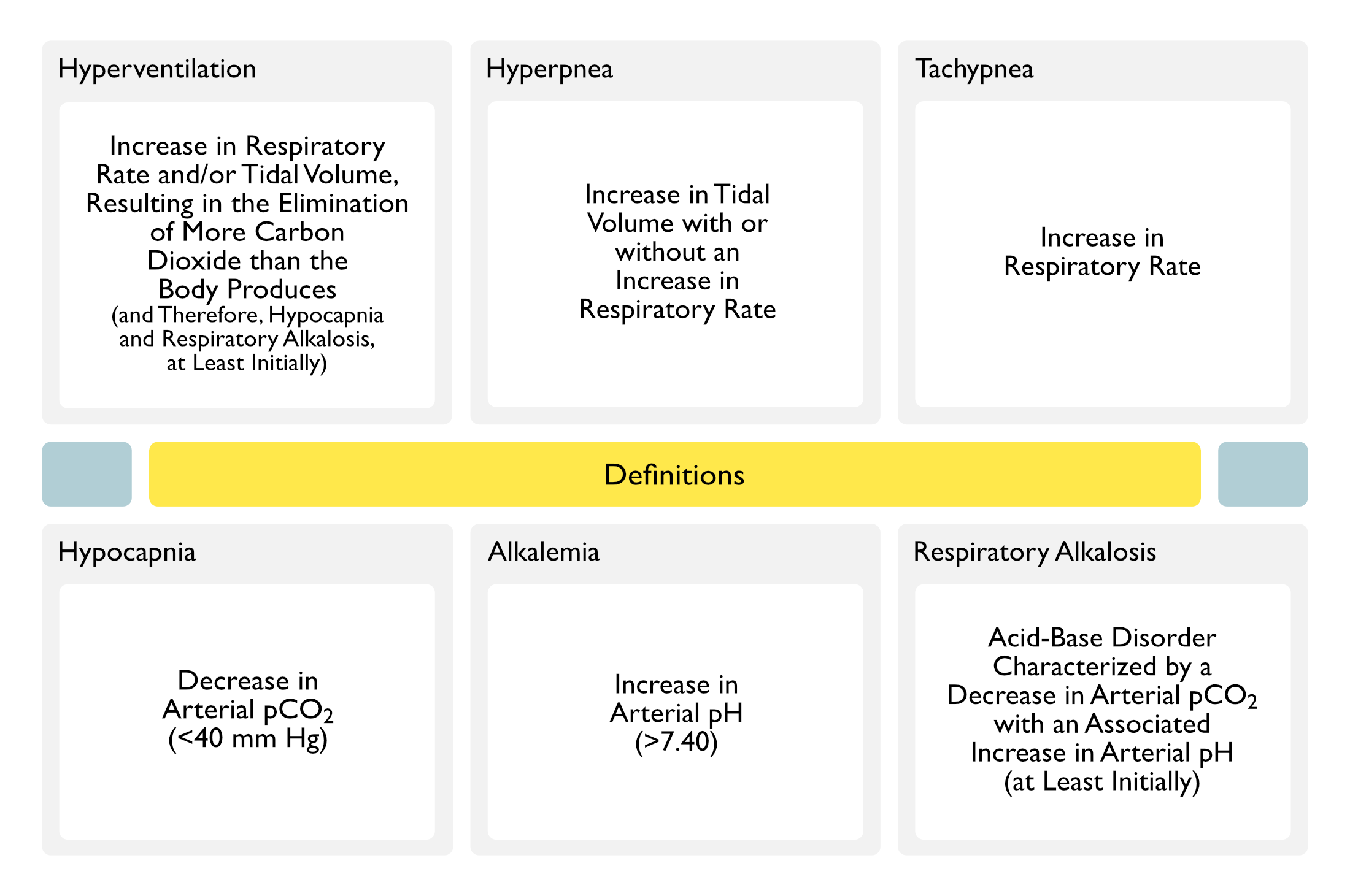
Hyperventilation is Defined as an Increase in Respiratory Rate and/or Tidal Volume, Resulting in the Elimination of More Carbon Dioxide than the Body Produces
Hyperventilation Results in Hypocapnia (Decreased Arterial pCO2) and Respiratory Alkalosis (at Least Initially, Until Renal Compensation Results in Bicarbonate Excretion with an Eventual Decrease in the pH Back Toward Normal) (see Respiratory Alkalosis)
Hypocapnia is Defined as Decrease in Arterial pCO2 (i.e. Decreased Arterial Blood Partial Pressure of Carbon Dioxide) to <40 mm Hg
Alkalemia
Definition
Alkalemia is Defined as Increase in the Arterial pH to >7.40
Note that a Patient Can Be Alkalemic without Having a Respiratory Alkalosis
Example
Metabolic Alkalosis Can Produce Alkalemia without the Presence of a Respiratory Alkalosis
Respiratory Alkalosis
Definition
Respiratory Alkalosis is Defined as an Acid-Base Disorder Characterized by a Decrease in Arterial pCO2 with an Associated Increase in Arterial pH (at Least Initially)
Note that a Patient Can Have a Respiratory Alkalosis without Being Alkalemic
Example
Due to Normal Compensatory Mechanisms, Chronic Respiratory Alkalosis Induces Metabolic (Predominantly Renal) Compensation (with a Progressive Decrease in Serum Bicarbonate Over Time), Culminating in Only Minimal/Absent Alkalemia
Hypoxemia-Induced Stimulation of Peripheral Chemoreceptors in the Carotid Bodies
However, the Degree of Hypoxemia-Induced Increase in Minute Ventilation is Modulated by Co-Existing pCO2 and pH, Mechanics of the Lung and Chest Wall, Genetic Factors, and the Overall Duration of Hypoxemia
The Increase in Minute Ventilation (VE) in Response to Decrease in pO2 is Non-Linear
The Most Pronounced Response is Observed with pO2 <60 mm Hg
The Increase in Minute Ventilation (VE) in Response to Decreased SaO2 is Relatively Linear
Clinical
On Mount Everest (Where pO2 is 27 mm Hg), the pCO2 is Decreased to 7.5 mm Hg (J Appl Physiol Respir Environ Exerc Physiol, 1983) [MEDLINE]
Hypoxemia-Induced Stimulation of Peripheral Chemoreceptors in the Carotid Bodies
However, the Degree of Hypoxemia-Induced Increase in Minute Ventilation is Modulated by Co-Existing pCO2 and pH, Mechanics of the Lung and Chest Wall, Genetic Factors, and the Overall Duration of Hypoxemia
The Increase in Minute Ventilation (VE) in Response to Decrease in pO2 is Non-Linear
The Most Pronounced Response is Observed with pO2 <60 mm Hg
The Increase in Minute Ventilation (VE) in Response to Decreased SaO2 is Relatively Linear
Pulmonary Disease (of Any Etiology Resulting in Hypoxemia)
Physiology
Hypoxemia-Induced Stimulation of Peripheral Chemoreceptors in the Carotid Bodies
However, the Degree of Hypoxemia-Induced Increase in Minute Ventilation is Modulated by Co-Existing pCO2 and pH, Mechanics of the Lung and Chest Wall, Genetic Factors, and the Overall Duration of Hypoxemia
The Increase in Minute Ventilation (VE) in Response to Decrease in pO2 is Non-Linear
The Most Pronounced Response is Observed with pO2 <60 mm Hg
The Increase in Minute Ventilation (VE) in Response to Decreased SaO2 is Relatively Linear
Stimulation of Peripheral and Central Chemoreceptors (and Increased Sensitivity of Peripheral Chemoreceptors to Hypoxia), Resulting in Increased Respiratory Drive
Sympathetic Overactivity, Increased Serum Progesterone and Vasoactive Intestinal Peptide (VIP), Increased Brain Ammonia and Glutamine, and Hypoxemia from the Formation of Small Intrapulmonary Shunts (ie: Hepatopulmonary Syndrome), All Resulting in Increased Respiratory Drive (Int J Cardiol, 2012) [MEDLINE]
Clinical
Severity of Respiratory Alkalosis Correlates with the Degree of Hepatic Insufficiency
Stimulation of Mechanical and Chemical Bronchopulmonary Receptors, Resulting in Increased Afferent Vagal Firing, Resulting in Increase in Respiratory Drive (and Cough and/or Bronchoconstriction)
Stimulation of Chest Wall Receptors (in Asthma, Pulmonary Fibrosis, and Chest Wall Disease), Resulting in Increased Respiratory Drive (and Dyspnea)
Stimulation of Mechanical and Chemical Bronchopulmonary Receptors, Resulting in Increased Afferent Vagal Firing, Resulting in Increase in Respiratory Drive (and Cough and/or Bronchoconstriction)
Stimulation of Mechanical and Chemical Bronchopulmonary Receptors, Resulting in Increased Afferent Vagal Firing, Resulting in Increase in Respiratory Drive (and Cough and/or Bronchoconstriction)
Stimulation of Mechanical and Chemical Bronchopulmonary Receptors, Resulting in Increased Afferent Vagal Firing, Resulting in Increase in Respiratory Drive (and Cough and/or Bronchoconstriction)
Renal Compensatory Response to Respiratory Alkalosis
In the Setting of Chronic Respiratory Alkalosis, the Kidney Both Decreases Acid Excretion (Which Results in a Positive Acid Balance that Decreases the Serum Bicarbonate Concentration) and Excretes Some Bicarbonate (Which Further reduces the Decreases the Serum Bicarbonate Concentration)
Mechanisms of Calcium Transport in the Blood (J Clin Invest, 1970) [MEDLINE] (Lancet, 1998) [MEDLINE]
Calcium Bound to Serum Proteins (Predominantly Albumin): 40-45%
Calcium Bound to Small Inorganic/Organic Anions (Phosphate, Citrate, Sulfate, Lactate, etc): 15%
Free (Ionized) Calcium: 40-45%
Ionized Calcium Concentration is Tightly Regulated by Parathyroid Hormone and Vitamin D
Clinical Manifestations in Acute Respiratory Alkalosis are More Common Than in Metabolic Alkalosis (see Metabolic Alkalosis)
Respiratory Alkalosis Probably Causes a Larger Change in Intracellular and Brain pH than Does Metabolic Alkalosis
Acute Respiratory Alkalosis Results in a Rapid Shift in Arterial pCO2, Which is Almost Immediately Transmitted Throughout the Total Body Water (Including the Intracellular Fluid Compartment, the Brain, and the Cerebrospinal Fluid)
This Accounts for the Characteristic Symptoms of Paresthesias, Carpopedal Spasm, and Lightheadedness Observed in Acute Respiratory Alkalosis
Metabolic Alkalosis-Associated Alterations in Blood Bicarbonate Cause Slower and Less Marked pH Changes within the Intracellular Fluid Compartment and Across the Blood-Brain Barrier
Cardiovascular Manifestations
Arrhythmias
Epidemiology
Hypocapnia has Been Linked to the Development of Arrhythmias, Both in Critically Ill Patients and in Patients with Panic Disorder
Physiology
Arrhythmias are Likely Mediated by Alkalemia-Mediated Electrophysiologic Effects on the Cardiac Conduction System (J Thorac Cardiovasc Surg, 1966) [MEDLINE]
May Also Be Mediated to Myocardial Ischemia (Although Specific Direct Myocardial Effects May Occur)
Treatment
Arrhythmias are More Resistant to Pharmacologic Treatment in the Setting of Alkalemia
In Traumatic Brain Injury, Prophylactic Hyperventilation is Associated with Worse Outcome (and is Therefore, Not Recommended)
Physiology
Decreased Cerebral Oxygenation
Although Hyperventilation May Transiently Decrease Intracranial Pressure, it May Do so at the Expense of Cerebral Perfusion
Additionally, Hypocapnia May Exacerbate Secondary Brain Injury, Because Increased Cerebral Vascular Reactivity and Vasoconstriction Can Result in Decreased Regional Cerebral Blood Flow
Reperfusion Injury
Rapid Correction of Severe Hypocapnia Causes Vasodilation in Ischemic Areas
In Ischemic Cerebrovascular Accident, Prophylactic Hyperventilation is Associated with Worse Outcome (and is Therefore, Not Recommended)
Physiology
Any Beneficial Effects of Hypocapnia on Intracranial Pressure are Likely Outweighed by the Effects of a Decreased Oxygen Supply (Due to Decreased Cerebral Perfusion)
Reperfusion Injury
Rapid Correction of Severe Hypocapnia Causes Vasodilation in Ischemic Areas
Postoperative Psychomotor Dysfunction
Epidemiology
Acute Hypocapnia is Common During General Anesthesia
Otherwise Healthy Patients Who are Subjected to Hypocapnia During General Anesthesia Have Been Found to Have Impaired Psychomotor Function for Up to 6 Days
Such Effects are Especially Pronounced in Older Patients
Seizures May Occur in the Setting of Hypocapnia (Lancet, 1998)[MEDLINE]
Physiology
Increased Neuronal Excitability, Seizure Activity, and Anaerobic Metabolism
Hypocapnic Potentiation of Seizure Activity, in Addition to Increasing Oxygen Demand, Augments Production of the Cytotoxic Excitatory Amino Acids Associated with Seizures
Hypocapnia May Also Induce Increased Neuronal Dopamine, Which May Increase the Risk of Seizures
Abnormal hyperventilation in patients with hepatic cirrhosis: role of enhanced chemosensitivity to carbon dioxide. Int J Cardiol. 2012 Jan 12;154(1):22-6. doi: 10.1016/j.ijcard.2010.08.066 [MEDLINE]
Physiology
Ionized calcium in normal serum, ultrafiltrates, and whole blood determined by ion-exchange electrodes. J Clin Invest. 1970;49(2):318 [MEDLINE]