Pisa Model (Am J Respir Crit Care Med, 2008) [MEDLINE]
Age
Age 57–67: coefficient 0.80 (Odds Ratio = 2.23)
Age 68–74: coefficient 0.87 (Odds Ratio = 2.38)
Age ≥75: coefficient 1.14 (Odds Ratio = 3.11)
Male Sex: coefficient 0.60 (Odds Ratio = 1.82)
Risk Factors
Immobilization: coefficient 0.42 (Odds Ratio = 1.53)
Deep Venous Thrombosis (Ever): coefficient 0.64 (Odds Ratio = 1.90)
Preexisting Diseases
Cardiovascular coefficient –0.51 (Odds Ratio = 0.60)
Pulmonary: coefficient –0.89 (Odds Ratio = 0.41)
Symptoms
Dyspnea (Sudden Onset): coefficient 2.00 (Odds Ratio = 7.38)
Orthopnea: coefficient –1.51 (Odds Ratio = 0.22)
Chest Pain: coefficient 1.01 (Odds Ratio = 2.74)
Fainting/Syncope: coefficient 0.66 (Odds Ratio = 1.93)
Hemoptysis: coefficient 0.93 (Odds Ratio = 2.52)
Signs
Unilateral Leg Swelling: coefficient 0.80 (Odds Ratio = 2.23)
Fever >38°C ( >100.4°F): coefficient –1.47 (Odds Ratio = 0.23)
Wheezes: coefficient –1.20 (Odds Ratio = 0.30)
Crackles: coefficient –0.61 (Odds Ratio = 0.54)
Electrocardiogram
Acute Cor Pulmonale (Includes ≥1 of the Following EKG Abnormalities: S1Q3T3, S1S2S3, Negative T-Waves in Right Precordial Leads, Transient Right Bundle Branch Block, or Pseudoinfarction Pattern): coefficient 1.96 (Odds Ratio = 7.11)
Constant: coefficient –3.43
Calculation
Add All of the Coefficients + Constant –3.43 to Obtain a Sum Score
Probability of Pulmonary Embolism = [1 + exp(–sum)]–1
Pulmonary Embolism Rule Out Criteria (PERC)
Rationale
Pulmonary Embolism Rule Out Criteria (PERC) were First Developed to Identify Patients with a Low Probability of Pulmonary Embolism in Whom the Risk of Unnecessary Diagnostic Testing Would Outweigh the Risk of Pulmonary Embolism (Ann Emerg Med, 2004) [MEDLINE]
Criteria
Age <50 y/o: 0 = meets criterion, 1 = does not meet criterion
Initial Heart Rate <100 bpm: 0 = meets criterion, 1 = does not meet criterion
Initial Room Air SaO2 >94%: 0 = meets criterion, 1 = does not meet criterion
Absence of Unilateral Leg Swelling: 0 = meets criterion, 1 = does not meet criterion
Absence of Hemoptysis: 0 = meets criterion, 1 = does not meet criterion
Absence of Surgery/Trauma within 4 wks: 0 = meets criterion, 1 = does not meet criterion
Absence of History of Venous Thromboembolism: 0 = meets criterion, 1 = does not meet criterion
Absence of Estrogen Use: 0 = meets criterion, 1 = does not meet criterion
Scoring
Pre-Test Probability with Score = 0 is <1%
Clinical Utility of Diagnostic Algorithms for the Diagnosis of Acute Pulmonary Embolism
Clinical Efficacy
Single-Center Study of the Use of Pulmonary Embolism Rule Out Criteria (PERC) in an Emergency Department (Ann Emerg Med, 2004) [MEDLINE]
Point of Care Pulmonary Embolism Rule Out Criteria (PERC) Doubled the Rate of Screening for Pulmonary Embolism in the Emergency Department
Point of Care Pulmonary Embolism Rule Out Criteria (PERC) Had a False-Negative Rate of <1%, Did Not Increase the Pulmonary Vascular Imaging Rate, and Decreased the Length of Stay
Prospective Observational Study of the Accuracy of Clinical Gestalt in the Diagnosis of Acute Pulmonary Embolism (Chest, 2005) [MEDLINE]
Accurate Determination of the Pretest Probability of Acute Pulmonary Embolism Appears to Increase with Clinical Experience
However, the Difference in Accuracy Between Inexperienced and Experienced Physicians is Not Sufficiently Large to Distinguish Between the Two When Determining Whether Clinical Gestalt or a Clinical Prediction Rule Should Be Used to Determine the Pretest Probability of Acute Pulmonary Embolism
Dutch Prospective Cohort Christopher Study Using Algorithm Combining Clinical Probability, D-Dimer, and Computed Tomography in the Diagnosis of Pulmonary Embolism (JAMA, 2006) [MEDLINE]: patients were followed for 3 mos
Initial Modified Wells Criteria Scoring: patients were categorized into “pulmonary embolism unlikely” vs “pulmonary embolism likely” groups
“Unlikely Group”: underwent D-dimer testing -> if D-dimer was normal, pulmonary embolism was considered excluded
All Others (“Likely” Group” or Those with Positive D-Dimer): underwent CT pulmonary angiogram to diagnose or exclude pulmonary embolism
Diagnostic Strategy Using Modified Wells Criteria, D-Dimer Testing, and CT Pulmonary Angiogram was Safe and Effective in the Diagnosis of Pulmonary Embolism
Prospective Multicenter Trial Examining the Effectiveness of Pulmonary Embolism Rule Out Criteria (PERC) in Patients at Low-Risk of Pulmonary Embolism ( J Thromb Haemost, 2008) [MEDLINE]: n = 8131 (85% of patients had a chief complaint of either dyspnea or chest pain), providers reported a low suspicion of pulmonary embolism in 20% of the patients
Low Pre-Test Probability for Pulmonary Embolism + PERC-Negative Had a Sensitivity of 97.4% and Specificity of 21.9%
The Combination of Gestalt Estimate of Low Pre-Test Probability for Pulmonary Embolism and PERC-Negative Decreases the Probability of Pulmonary Embolism to <2% in About 20% of Outpatients with Suspected Pulmonary Embolism
Secondary Analysis of a Prospective Database Examining the Effectiveness of Pulmonary Embolism Rule Out Criteria (PERC) in the Emergency Deopartment (Am J Emerg Med, 2008) [MEDLINE]
Pulmonary Embolism Rule Out Criteria (PERC) Had 100% Sensitivity and 16% Specificity
Pulmonary Embolism Rule Out Criteria (PERC) Had 14% Positive Predictive Value and 100% Negative Predictive Value
Dutch (Prometheus Study Group) Prospective Cohort Study of Clinical Decision Rules in the Diagnosis of Pulmonary Embolism (Ann Intern Med, 2011) [MEDLINE]
All 4 Clinical Decision Rules (Wells Rule, Revised Geneva Score, Simplified Wells Rule, and Simplified Revised Geneva Score) Had Similar Performance for the Exclusion of Acute Pulmonary Embolism in Combination with a Normal D-Dimer Result
Retrospective Study of the Effectiveness of Pulmonary Embolism Rule Out Criteria (PERC) in the Emergency Department (J Thromb Haemost, 2011) [MEDLINE]
Pulmonary Embolism Rule Out Criteria (PERC) is Only Valid in Patients with a Low Pre-Test Probability of Pulmonary Embolism (<15%): the predictive value is significantly lower in patients with intermediate/high pre-test probability for pulmonary embolism and should not be used in these populations
Systematic Review and Meta-Analysis of Pulmonary Embolism Rule Out Criteria (PERC) in Acute Pulmonary Embolism (Emerg Med J, 2013) [MEDLINE]
Overall Proportion of Missed Pulmonary Emboli by Using PERC was Only 0.3%
PERC Had 97% Sensitivity and 22% Specificity: indicates that 22% of D-dimer tests could have been safely avoided had the PERC been universally applied
Dutch Study of the Accuracy of Clinical Decision Rules in the Diagnosis of Acute Pulmonary Embolism in Patients with a Delayed Clinical Presentation (Am J Respir Crit Care Med, 2013) [MEDLINE]
Pulmonary Embolism Can Be Safely Excluded Based on Clinical Decision Rules and D-Dimer Testing in Patients with a Delayed Clinical Presentation
Patients with a Delayed Clinical Presentation More Frequently Had a Centrally-Located Pulmonary Embolism
Cumulative Rates of Recurrent Venous Thromboembolism and Mortality Were Not Different for Patients with and without a Delayed Clinical Presentation
European ADJUST-PE Study of Age-Adjusted D-Dimer Levels in the Diagnosis of Pulmonary Embolism (JAMA, 2014) [MEDLINE]
Age-Adjusted D-Dimer (Only for Patients ≥50 y/o): defined as 10 x age
Compared with a Fixed D-Dimer Cutoff of 500 μg/L (500 ng/mL), the Combination of a Pre-Test Clinical Probability Assessment and Age-Adjusted D-Dimer Cutoff was Associated with a Larger Number of Patients in Whom Pulmonary Embolism Could Be Considered Ruled Out with a Low Likelihood of Subsequent Clinical Venous Thromboembolism
Systematic Review and Meta-Analysis of Wells Criteria and D-Dimer Testing in the Diagnosis of Pulmonary Embolism (Ann Intern Med, 2016) [MEDLINE]
In Patients with an “Unlikely” Pre-Test Probability of Pulmonary Embolism, Age-Adjusted D-Dimer Testing is Associated with a 5% Increase in the Proportion of Patients with Suspected Pulmonary Embolism in Whom Imaging Can Be Safely Withheld, as Compared to Fixed D-Dimer Testing
Study of the Yield of CT Pulmonary Artery Angiogram When Providers Used Well Criteria-Based Clinical Decision Support in the ED (Radiology, 2016) [MEDLINE]
Odds of Diagnosis of Acute Pulmonary Embolism was 2x Higher (Yield: 11.2% of Studies) When Providers Adhered to Wells Criteria-Based Clinical Decision Support, as Compared to Providers who Overrode the Clinical Decision Support (Yield: 4.2% of Studies)
Wells Criteria ≤4: providers were recommended to initially use D-dimer testing (since the probability of acute PE is low in this population, if D-dimer level is normal)
Providers Who Overrode the Clinical Decision Support Ordered a CT Pulmonary Angiogram in Patients with Wells Criteria ≤4 and Negative D-Dimer (or Did Not Perform D-Dimer Testing)
If the Pre-D-Dimer Probability of PE is 15% (Intermediate Pre-Test Probability), Only a D-DImer <500 ng/mL Will Result in a Post-Test Probability <3%
Consequently, Given a Pre-Test Probability of 15% (Intermediate Pre-Test Probability) and a CT Pulmonary Artery Angiogram Threshold of 3%, a Strategy to Obtain CT Pulmonary Artery Angiogram for D-Dimer ≥500 ng/ mL is Consistent with the Interval Likelihood Ratios Reported
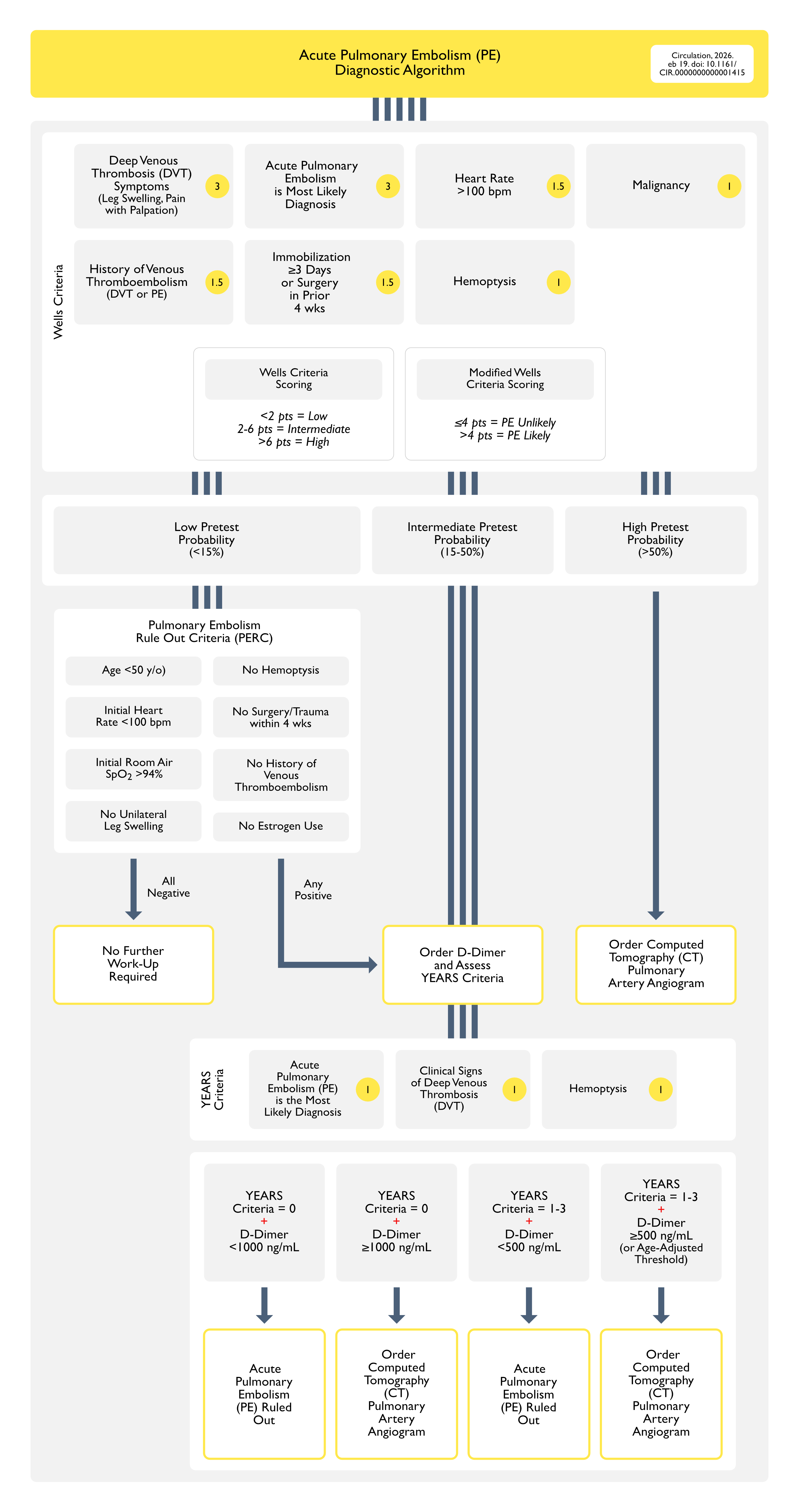
Dutch Multicenter Prospective Cohort YEARS Study (Lancet, 2017) [MEDLINE]: n= 3616
D-Dimer and Three Years Items were Assessed (Clinical Signs of Deep Venous Thrombosis, Hemoptysis, Pulmonary Embolism as the Most Likely Diagnosis
YEARS Diagnostic Algorithm Safely Excluded the Diagnosis of Pulmonary Embolism
Years Algorithm Resulted in an Absolute 14% Decrease of CT Pulmonary Artery Angiogram Studies in All Ages and Across Several Relevant Subgroups
French Randomized PROPER Trial Using Pulmonary Embolism Rule-Out Criteria (PERC) in Low-Risk Emergency Department Patients (JAMA, 2018) [MEDLINE]: crossover cluster-randomized clinical noninferiority trial in 14 emergency departments in France
In Patients at Very Low-Risk with Suspected Pulmonary Embolism, PERC Strategy vs Conventional Strategy Had Similar Rates of Thromboembolic Events Over 3 mo
PERC Strategy is Safe for Use in Very Low-Risk Patients Presenting to the Emergency Department
Recommendations (American College of Physicians Guidelines for the Evaluation of Patients with Suspected Pulmonary Embolism, 2015) (Ann Intern Med, 2015) [MEDLINE]
Use Validated Clinical Prediction Rules (or Clinical Gestalt) to Estimate the Pretest Probability of Pulmonary Embolism
Clinical Gestalt
The Overall Accuracy of an Experienced Clinician’s Gestalt Appears to Be Similar to that of Clinical Decision Rules
However, Clinical Decision Rules Allow Standardization for Evaluation of Suspected Acute Pulmonary Embolism by Clinician Who Less Frequently Evaluate Patients with Suspected Acute Pulmonary Embolism
Clinical Decision Rules
Wells Criteria or Modified (Dichotomized) Wells Criteria
Revised Geneva Score or Revised Simplified Geneva Score
Kline Rule
Pisa Model
Low Pre-Test Probability of Pulmonary Embolism + Meets All Pulmonary Embolism Rule Out Criteria (PERC)
D-Dimer and Imaging Studies are Not Recommended
Intermediate Pre-Test Probability of Pulmonary Embolism + Does Not Meet All Pulmonary Embolism Rule Out Criteria (PERC)
Initial High-Sensitivity D-Dimer (But Not Imaging Study) is Recommended as the Initial Diagnostic Test
Use Age-Adjusted D-Dimer Thresholds (Age x 10) Rather than Generic 500 ng/mL Threshold in Patients >50 y/o
If D-Dimer is Below the Age-Adjusted Cutoff, Do Not Use Imaging Study
High Pre-Test Probability of Pulmonary Embolism
CT Pulmonary Artery Angiogram (But Not D-Dimer) is Recommended
Ventilation/Perfusion (V/Q) Scan Should Be Reserved for Patients with Contraindication to CT Pulmonary Artery Angiography (or When CT Pulmonary Artery Angiography is Not Available)
Recommendations (European Society of Cardiology and European Respiratory Society Guidelines for the Diagnosis and Management of Acute Pulmonary Embolism, 2019) (Eur Heart J, 2020) [MEDLINE]
For Suspected Acute Pulmonary Embolism without Hemodynamic Instability, Use of Validated Criteria for Diagnosing Acute Pulmonary Embolism is Recommended (Class I, Level B)
It is Recommended that the Diagnostic Strategy Be Based on Clinical Probability (Assessed Either by Clinical Judgement or By a Validated Prediction Rule (Class I, Level A)
Recommendations (Consensus Practice from the PERT Consortium, 2019) (Clin Appl Thromb Hemost, 2019) [MEDLINE]
Use a Combination of Low or Intermediate Pretest Probability, the PERC Rule, and D-Dimer Testing to Rule Out Acute Pulmonary Embolism without Imaging
When Possible, Use CT Pulmonary Artery Angiogram to Diagnose Acute Pulmonary Embolism in Patients with Low or Intermediate Pretest Probability and a Positive D-Dimer, or High Pretest Probability
Echocardiography and/or Portable V/Q Scan (When Available), Should Be Considered When There are Contraindications to or Inability to Obtain CT Pulmonary Artery Angiogram
Additionally, Duplex Ultrasonography Should Be Considered to Confirm the Presence, Acuity, and Extent of Venous Thromboembolism
Recommendations (2026 AHA/ACC/ACCP/ACEP/CHEST/SCAI/SHM/SIR/SVM/SVN Guideline for the Evaluation and Management of Acute Pulmonary Embolism in Adults: A Report of the American College of Cardiology/American Heart Association Joint Committee on Clinical Practice Guidelines) (Circulation, 2026) [MEDLINE]
In Patients Presenting with Symptoms Suggestive of Acute Pulmonary Embolism (PE), a Targeted History and Comprehensive Physical Examination is Recommended to Assist in Determining the Clinical Pretest Probability of Acute Pulmonary Embolism (PE) (Class of Recommendation: 1; Level of Evidence: A)
In Adult Patients Undergoing Evaluation for Pulmonary Embolism (PE) and Who Have a Low/Intermediate Clinical Probability of Pulmonary Embolism (PE) (<50%) by Risk Assessment, an Age-Adjusted D-Dimer Value Below the Threshold (Age × 10 μg/L for Fibrinogen Equivalent Units Assays) Effectively Excludes Pulmonary Embolism (PE) and the Need for Imaging (Class of Recommendation: 2a; Level of Evidence: B-R)
In Adult Patients with Suspected Pulmonary Embolism (PE), the YEARS Algorithm Can Be Useful to Identify Which Patients Do Not Need Imaging to Rule Out Pulmonary Embolism (PE) (Class of Recommendation: 2a; Level of Evidence: B-R)
YEARS Algorithm
In Patients Who Have ≥1 of the Following YEARS Criteria, Use a D-Dimer Threshold of 500 μg/L
Acute Pulmonary Embolism (PE) as the Most Likely Diagnosis
Clinical Signs of Deep Venous Thrombosis (DVT)
Hemoptysis
In Patients Who Have for Have No YEARS Criteria, Use a D-Dimer Threshold of 1000 μg/L
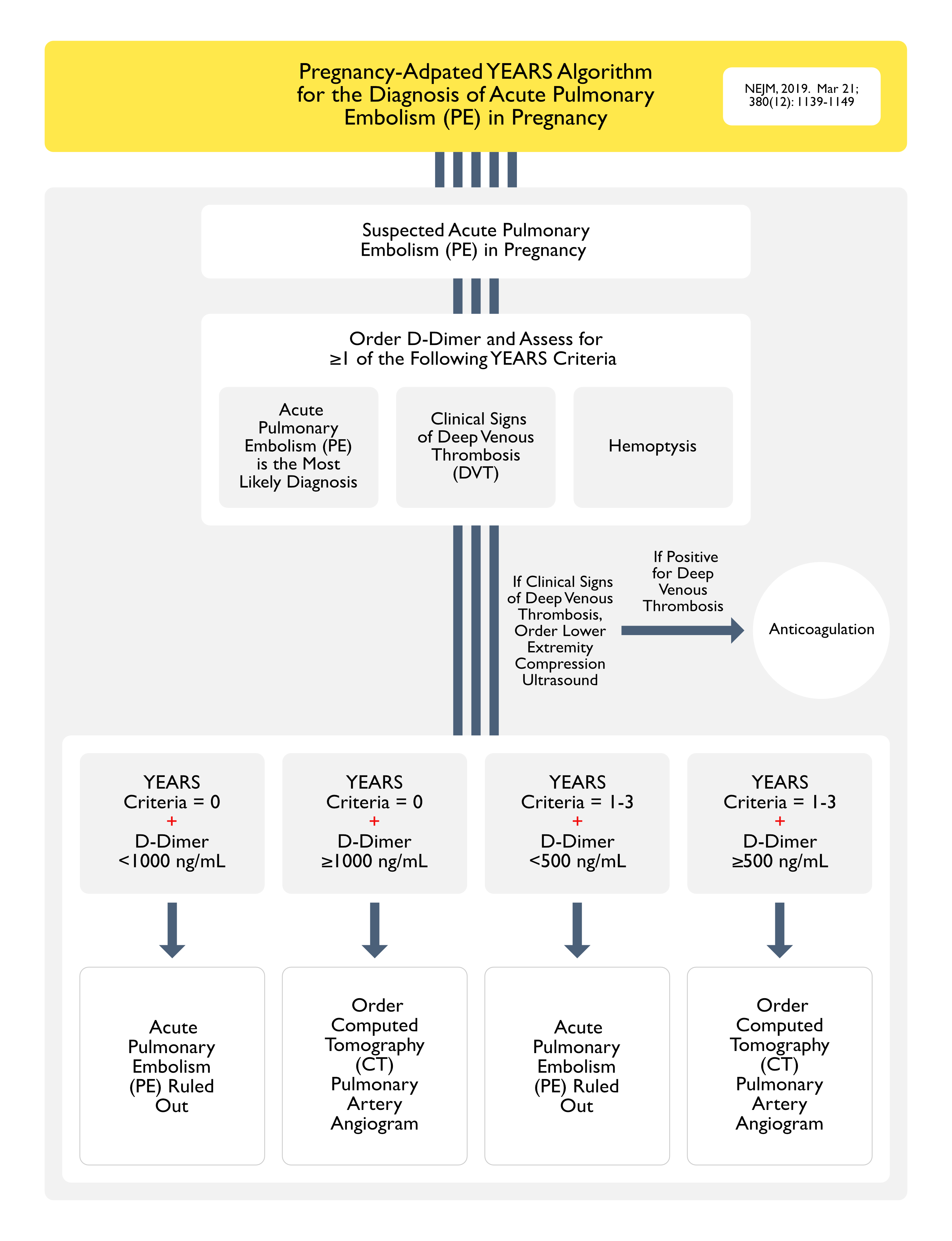
In Pregnant Adults, it May Be Reasonable to Use Pregnancy-Adapted YEARS Criteria to Identify Patients Who Do Not Need Imaging for Pulmonary Embolism (PE) (Class of Recommendation: 2b; Level of Evidence: B-R) (NEJM, 2019) [MEDLINE]
In Patients Presenting with Symptoms/Signs Suggestive of Acute Pulmonary Embolism (PE), Imaging is Recommended for Those Who are Deemed High Probability (>50% Probability of Pulmonary Embolism) by a Validated Clinical Risk Prediction Score or an Elevated D-Dimer Level in Order to Confirm/Exclude Pulmonary Embolism (PE) (Class of Recommendation: 1; Level of Evidence: A)
In Patients Undergoing Imaging Evaluation for Suspected Acute Pulmonary Embolism (PE), a Positive Computed Tomography (CT) Pulmonary Artery Angiogram or High Probability Ventilation/Perfusion (V/Q) Scan is Sufficient to Diagnose Pulmonary Embolism (PE) (Class of Recommendation: 1; Level of Evidence: A)
In Patients Undergoing Imaging Evaluation, a Computed Tomography (CT) Pulmonary Artery Angiogram is Recommended in Preference to a V/Q Scan to Confirm the Diagnosis of Acute Pulmonary Embolism (PE) (Class of Recommendation: 1; Level of Evidence: B-R)
In Pregnant Patients Presenting with Symptoms, YEARS Criteria Suggestive of Acute Pulmonary Embolism (PE), and a Normal Chest X-Ray, Imaging Evaluation with Low-Radiation Dose Computed Tomography (CT) Pulmonary Artery Angiogram is Reasonable Over Low-Dose Perfusion Scintigraphy (Class of Recommendation: 2a; Level of Evidence: B-NR)
In Patients with Suspected Acute Pulmonary Embolism (PE) Who Cannot Undergo a Computed Tomography (CT) Pulmonary Artery Angiogram, it is Reasonable to Perform a V/Q Scan in Preference to Contrast-Enhanced Magnetic Resonance Angiography (MRA) to Improve the Diagnostic Yield (Class of Recommendation: 2a; Level of Evidence: B-R)
In Patients with Confirmed Acute Pulmonary Embolism (PE), Obtaining Lower Extremity Venous Duplex Ultrasound Examination May Be Reasonable in Patients with Clinical Findings Suggestive of Deep Venous Thrombosis (DVT) or if the Presence of Deep Venous Thrombosis (DVT) Will Change Management or Inform Prognosis (Class of Recommendation: 2b; Level of Evidence: B-NR)
Clinical Grading/Risk Stratification of Pulmonary Embolism Severity
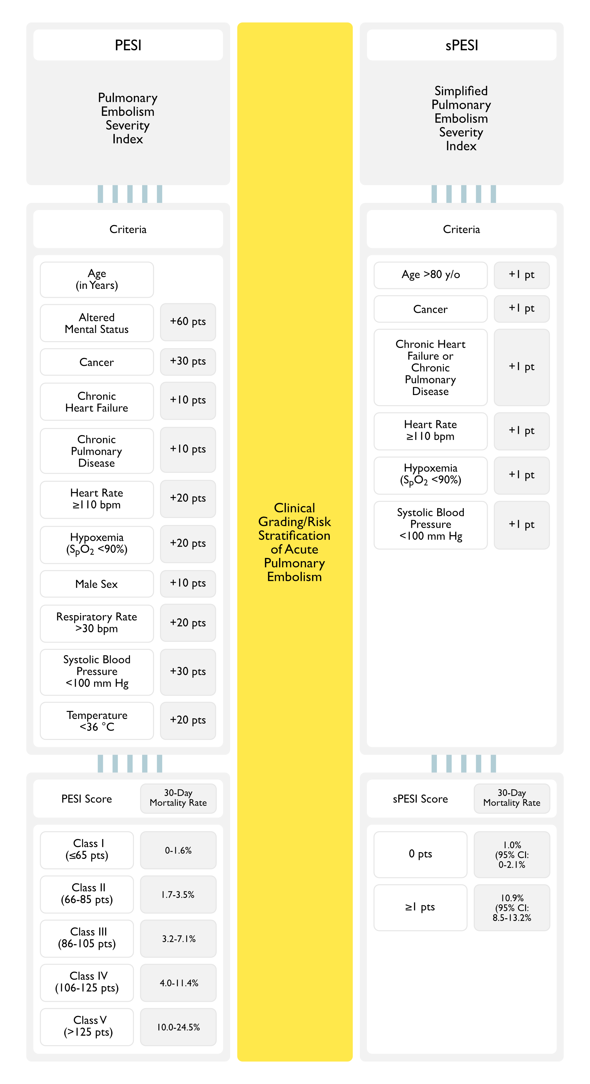
Pulmonary Embolism Severity Index (PESI) (Am J Respir Crit Care Med, 2005) [MEDLINE]
Simplified Pulmonary Embolism Severity Index (sPESI)
Simplified Pulmonary Embolism Severity Index (sPESI) Has Similar Prognostic Accuracy and Clinical Utility and Greater Ease of Use, as Compared with the Original Pulmonary Embolism Severity Index (PESI)
Criteria
Age >80 y/o: 1 pt
Cancer: 1 pt
Chronic Heart Failure or Chronic Pulmonary Disease: 1 pt
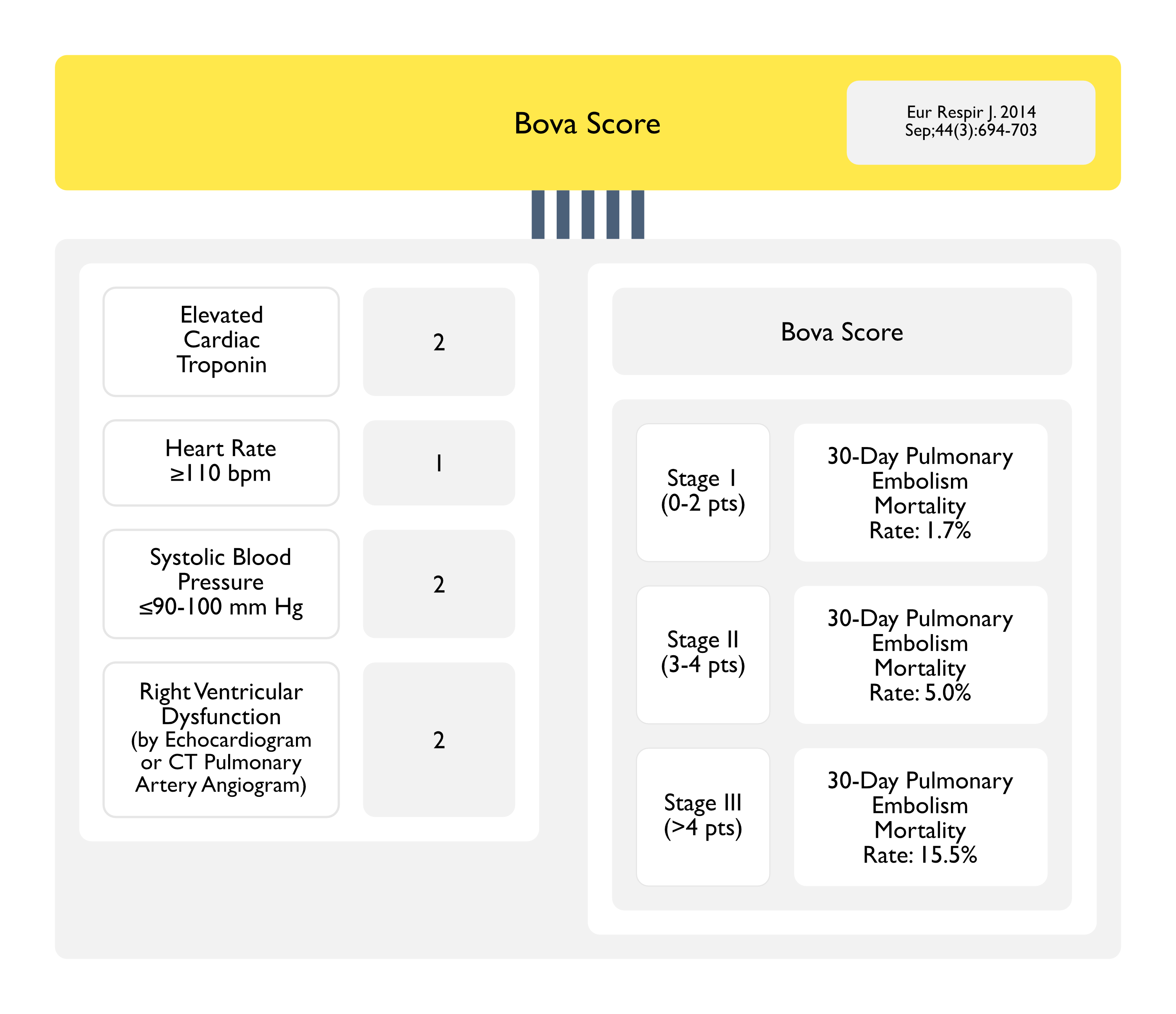
Retrospective Validation Study of Bova Score for the Identification of Normotensive Acute Symptomatic Pulmonary Embolism Patients at Intermediate-High Risk for Complications (Chest, 2015) [MEDLINE]: n = 1,083
The Bova Score Classified the Majority of the Cohort into the Lowest Bova Risk Stage (Stage I, 80%; Stage II, 15%; Stage III, 5%)
The Primary End Point Occurred in 8.4% of the Patients (95% CI: 6.7%-10%) During the 30 Days After Pulmonary Embolism Diagnosis
Risk Stage Correlated with the Pulmonary Embolism-Related Complication Rate (Class I: 4.4%; Class II: 18%; Class III: 42%; ICC 0.93 [95% CI, 0.92-0.94]; κ Statistic, 0.80; P < .001), In-Hospital Complication Rate (Class I: 3.7%; Class II: 15%; Class III: 37%), and 30-Day Pulmonary Embolism-Related Mortality (Class I: 3.1%; Class II: 6.8%; Class III: 10.5%)
Prospective Validation of Bova Score in Normotensive Patients with Acute Pulmonary Embolism (Thromb Res, 2018) [MEDLINE]: n = 639
Bova Risk Stage Correlated with Acute Pulmonary Embolism Complication Rate
Stage I: 2.9%
Stage II: 17%
Stage III: 27%
Patients Classified as Stage III Had a 6.5-Fold Increase in Risk for Adverse Outcomes (3.1-13.5, p < 0.001), as Compared with Stages I and II Combined
Rescue Thrombolysis Increased from Stage I to Stage III (0.6%, 12% and 15%, Respectively)
However, All-Cause Mortality (5.3%) Did Not Significantly Differ Among the Stages
Systematic Review and Meta-Analysis of Bova Score in Acute Pulmonary Embolism (PE) (Thromb Res, 2020) [MEDLINE]: n = 8,342 (from 9 studies)
Number of Patients in Each Class
Class I: 71.4%
Class II: 20.2%
Class III: 8.4%
Incidence of Short-Term Pulmonary Embolism-Related Composite Adverse Outcome
As Compared with Risk Class I and II, Class III was Significantly Associated with Short-Term Pulmonary Embolism-Related Composite Adverse Outcome (Odds Ratio: 5.45, 95% CI, 3.70-8.02) and Pulmonary Embolism-Related Mortality (Odds Ratio: 5.09, 95% CI, 3.54-7.30)
Pooled Sensitivity, Specificity, Positive Likelihood Ratio and Negative Likelihood Ratio of the Score for Predicting Short-Term Composite Adverse Outcome were 0.25 (95% CI: 0.22-0.29), 0.93 (95% CI: 0.92-0.93), 4.05 (95% CI: 2.90-5.67) and 0.81 (95% CI: 0.74-0.88), Respectively
The Weighted Area Under the Summarized Receiver Characteristics Operation Curve for Predicting Composite Adverse Outcome was 0.73 ± 0.09
Hestia Criteria
Criteria
Is the Patient Hemodynamically Unstable?
Is Thrombolysis or Embolectomy Required?
Does the Patient Have Active Bleeding or a High Risk of Bleeding?
Does the Patient Require >24 hrs of Outpatient Oxygen to Maintain Oxygen Saturation >90%?
Is Pulmonary Embolism Diagnosed During Anticoagulant Treatment?
Does the Patient Have Severe Pain Requiring Intravenous Pain Medication for >24 hrs?
Are There Medical/Social Reasons for Hospitalization >24 hrs (Such as Infection, Cancer, or Lack of Support System)?
Does the Patient Have a Creatinine Clearance <30 mL/min?
Does the Patient Have Severe Liver Impairment?
Is the Patient Pregnant?
Does the Patient Have a Documented History of Heparin-Induced Thrombocytopenia?
In Retrospective Studies, Approximately 40% of Patients with Intermediate-Risk Acute Pulmonary Embolism (PE) Had Decreased Cardiac Index Consistent with Normotensive Cardiogenic Shock (Catheter Cardiovasc Interv, 2020) [MEDLINE]
Note that the Concept of Normotensive Shock Has Been Derived from Retrospective Observational Data in a Specific Subset of Patients Who Underwent Mechanical Thrombectomy (Not Data from Randomized Prospective Trials of All Patients with Pulmonary Embolism) (JACC Cardiovasc Interv, 2023) [MEDLINE]
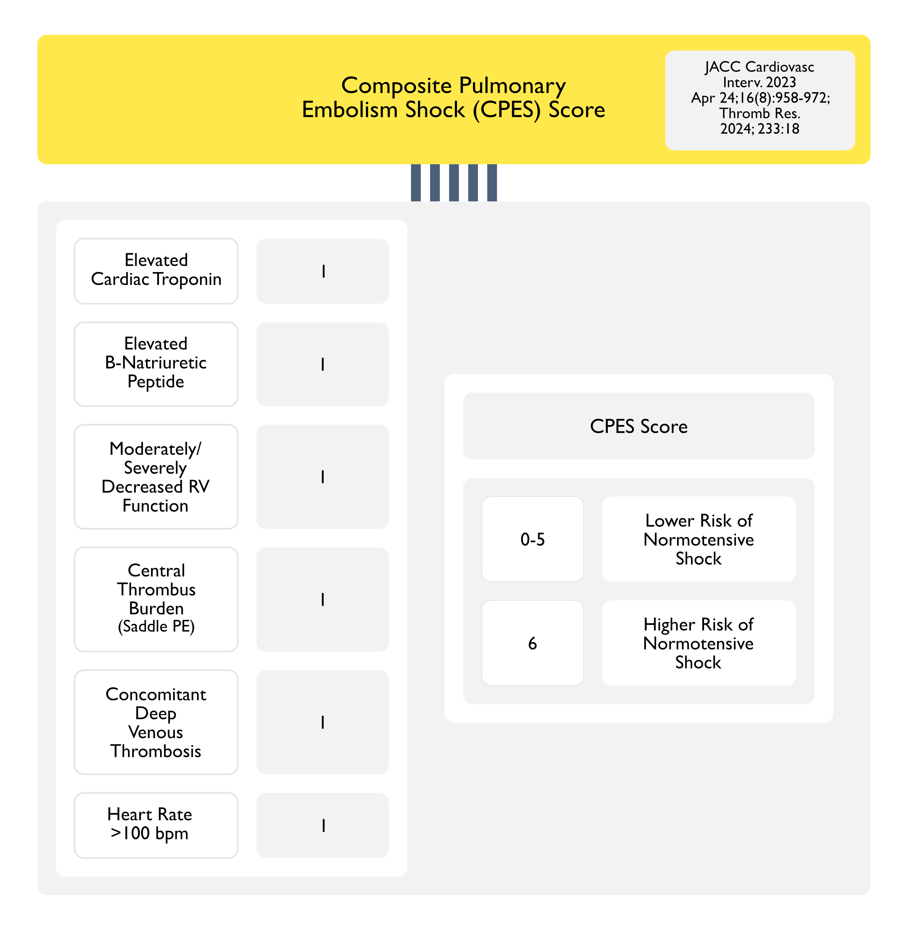
Composite Pulmonary Embolism Shock (CPES) Score was Developed to Identify Pulmonary Embolism Patients with Normotensive Shock (Thromb Res, 2024) [MEDLINE]
In Hemodynamically-Stable Acute Pulmonary Embolism
CPES ≤3 is Associated with a 30-Day All-Cause Mortality Rate of 4.2%
CPES >3 is Associated with a 30-Day All-Cause Mortality Rate of 7.7%
Criteria
Elevated Cardiac Troponin: 1 pt
Elevated B-Natriuretic Peptide (BNP): 1 pt
Moderately/Severely Decreased Right Ventricular Function: 1 pt
Central Thrombus Burden (Saddle PE): 1 pt
Concomitant Deep Venous Thrombosis (DVT): 1 pt
Heart Rate >100 bpm: 1 pt
Scoring
Score 0-5: Lower Risk of Normotensive Shock
Score 6: Higher Risk of Normotensive Shock
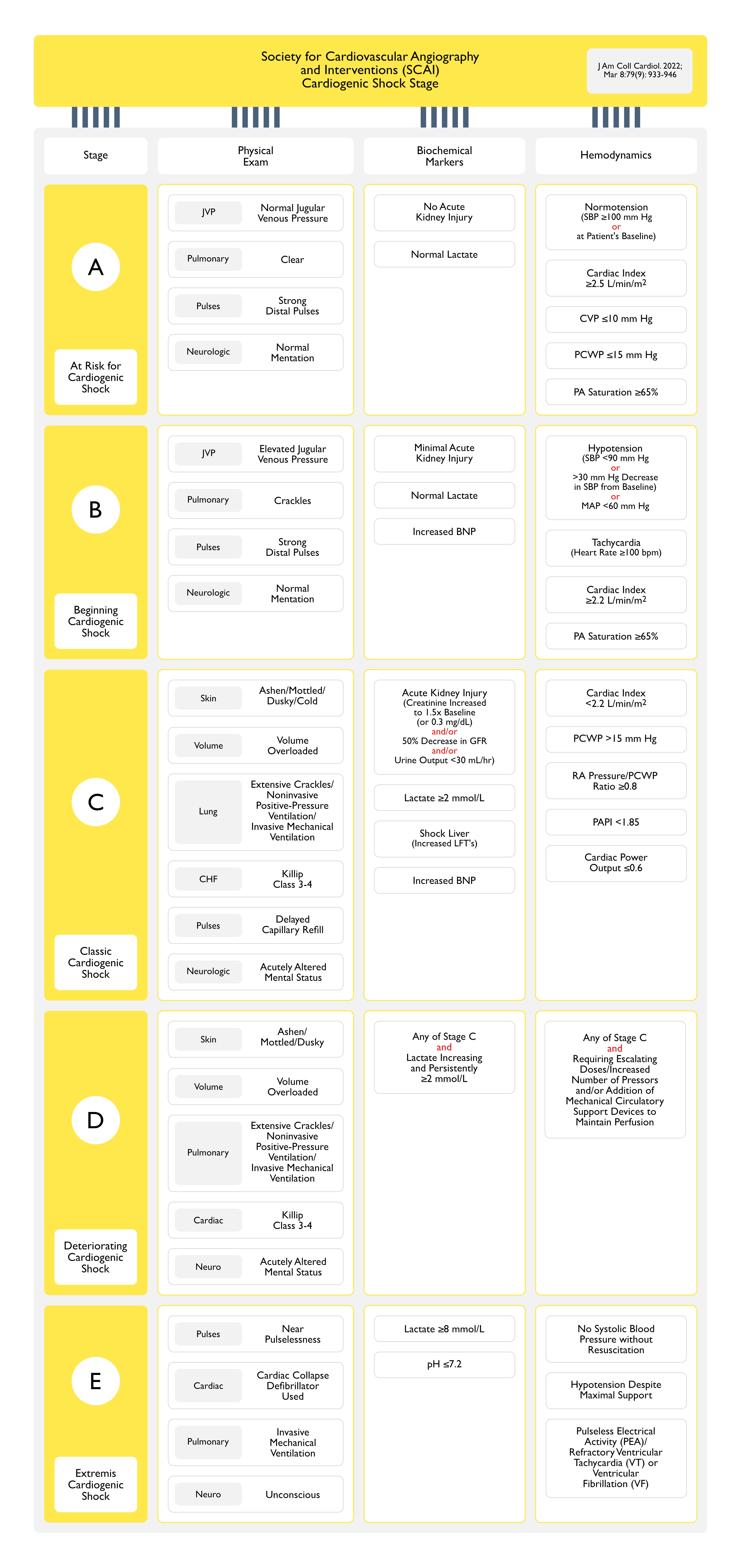
Society for Cardiovascular Angiography and Interventions (SCAI) Cardiogenic Shock Stage (J Am Coll Cardiol, 2022) [MEDLINE]
Shock Index
Criteria
Heart Rate/Systolic Blood Pressure
Scoring
Lower Scores Associated with Lower Risk
There is a Lack of Consensus on Which Cut-Points to Use for Pulmonary Embolism Risk Stratification
Other Clinical Grading/Risk Stratification Criteria
Clinical Data
Grading of CT Signs of Right Ventricular Dysfunction in Acute Pulmonary Embolism (AJR Am J Roentgenol, 2010) [MEDLINE]
Volumetric Determination of the Right Ventricular Volume/Left Ventricular Volume Ratio
Ratio >1.2 is Suggestive of Right Ventricular Strain
Most Reproducible/Least User-Dependent of the CT Measurements (as Compared to Septal Bowing or IVC Reflux)
Grading of Factors Associated with 30-Day Frequency of Adverse Events in Prep Study (Am J Respir Crit Care Med, 2010) [MEDLINE]
Altered Mental Status: OR 6.8 (95% CI: 2.0-23.3)
Shock on Admission: OR 2.8 (95% CI: 1.1-7.5)
Cancer: OR 2.9 (95% CI: 1.2-6.9)
Elevated BNP: OR 1.3 for an increase of 250 ng/L (95% CI: 1.1-1.6)
Echocardiographic Right Ventricular Volume/Left Ventricular Volume Ratio: OR 1.2 for an increase of 0.1 (95% CI: 1.1-1.4)
Grading of Pulmonary Embolism Using Right Ventricular Dysfunction and Troponin Levels (Chest, 2013) [MEDLINE]
Right Ventricular Dysfunction and Elevated Troponin Level: these criteria have an incremental prognostic value for risk stratification in hemodynamically-stable patients with acute pulmonary embolism
Recommendations (European Society of Cardiology and European Respiratory Society Guidelines for the Diagnosis and Management of Acute Pulmonary Embolism, 2019) (Eur Heart J, 2020) [MEDLINE]
Initial Risk Stratification of Suspected/Confirmed Acute Pulmonary Embolism (Based on the Presence of Hemodynamic Instability) is Recommended to Identify Patients at High Risk of Early Mortality (Class I, Level B)
Low-Risk Acute Pulmonary Embolism
Defined as Acute Pulmonary Embolism with Hemodynamic Stability and Absence of Right Ventricular Dilation/Dysfunction and/or Positive Biomarkers Suggestive of Myocardial Injury/Myocardial Distention
30-Day Mortality Rate was 0.5% (95% CI: 0-1.0; Negative Predictive Value 99.5%) (Eur Respir J, 2016) [MEDLINE]
Defined as Acute Pulmonary Embolism with Class III-V PESI ≥86 pts (or sPESI ≥1 pt) or Right Ventricular Dilation/Dysfunction (by CT PA Angiogram or Echocardiography), or Elevated Serum Troponin
Note: Elevation of Other Laboratory Biomarkers (Such as NT-ProBNP ≥600 ng/L, H-FABP ≥6 ng/mL, or Copeptin ≥24 pmol/L) May Provide Additional Prognostic Information (However, These Biomarkers Have Been Validated in Cohort Studies, But They Have Not Been Used to Guide Treatment Decisions in Randomized Controlled Trials)
Subclassification Per the European Society of Cardiology and European Respiratory Society Guidelines (Eur Heart J, 2020) [MEDLINE]
Intermediate-Low Risk: presence of either right ventricular dysfunction or troponin elevation (30-Day Mortality Rate was 6.0%; 95% CI: 3.4-8.6) (Eur Respir J, 2016) [MEDLINE]
Intermediate-High Risk: presence of both right ventricular dysfunction and troponin elevation (with any PESI/sPESI score) (30-Day Mortality Rate was 7.7%; 95% CI: 4.5-10.9) (Eur Respir J, 2016) [MEDLINE]
High-Risk (“Massive”) Acute Pulmonary Embolism
Defined as Acute Pulmonary Embolism with Hemodynamic Instability Characterized by Any of the Following
Cardiac Arrest
Obstructive Shock (Systolic Blood Pressure <90 mm Hg or Vasopressors Required to Achieve a Blood Pressure ≥90 mmHg Despite Adequate Filling Status, in Combination with End-Organ Hypoperfusion)
Persistent Hypotension (Systolic Blood Pressure <90 mm Hg or a Systolic Blood Pressure Decrease ≥40 mm Hg for >15 min, Not Caused by New-Onset Arrhythmia, Hypovolemia, or Sepsis)
The Presence of Remaining Variables (PESI Score ≥86 pts/sPESI ≥1/Elevated Troponin/etc) are Not Required to Define a Patient as High-Risk
30-Day Mortality Rate was 22% (95% CI: 14.0-29.8)
In Patients without Hemodynamic Instability, Further Stratification of Patients with Acute Pulmonary Embolism into Low and Intermediate Risk Categories is Recommended (Class I, Level B)
In Patients without Hemodynamic Instability, Use of Clinical Prediction Rules Integrating Acute Pulmonary Embolism Severity and Comorbidity (Preferably the PESI or sPESI) Should Be Considered for Risk Assessment in the Acute Phase of Pulmonary Embolism (Class IIa, Level B)
Assessment of the Right Ventricle by Imaging Methods or Laboratory Biomarkers (Troponin or Brain Natriuretic Peptide) Should Be Considered, Even in the Presence of a Low PESI or a Negative sPESI (Class IIa, Level B)
In Patients without Hemodynamic Instability, Use of Validated Scores Combining Clinical, Imaging, and Laboratory Acute Pulmonary Embolism-Related Prognostic Factors May Be Considered to Further Stratify the Severity of the Acute Pulmonary Embolism Episode (Class IbI, Level C)
Recommendations (Consensus Practice from the PERT Consortium, 2019) (Clin Appl Thromb Hemost, 2019) [MEDLINE]
Once Acute Pulmonary Embolism is Diagnosed, Risk Stratification is Recommended Using a Composite of Clinical Appearance, Systolic Blood Pressure, Heart Rate, Respiratory Rate, Oxygen Requirement, PESI or sPESI, Imaging for Right Ventricular Dysfunction (by CT PA Angiogram or Echocardiography) and/or Biomarkers (Troponin, BNP, or NT-pro-BNP)
Recommendations (2026 AHA/ACC/ACCP/ACEP/CHEST/SCAI/SHM/SIR/SVM/SVN Guideline for the Evaluation and Management of Acute Pulmonary Embolism in Adults: A Report of the American College of Cardiology/American Heart Association Joint Committee on Clinical Practice Guidelines) (Circulation, 2026) [MEDLINE]
Clinical Risk Scoring Systems
In Patients Diagnosed with Acute Pulmonary Embolism (PE) in AHA/ACC PE Categories A and B, Use of the Hestia, PESI, and/or sPESI Risk Scores is Recommended to Identify Patients with a Low Risk for Short-Term Adverse Outcomes (Class of Recommendation: 1; Level of Evidence: B-R)
In Hemodynamically Stable Patients Diagnosed with Acute Pulmonary Embolism (PE) in AHA/ACC Pulmonary Embolism Categories C and D, Using a Validated Pulmonary Embolism-Specific Risk Score is Reasonable to Identify Patients with a Higher Risk for Short-Term Adverse Outcomes (Class of Recommendation: 2a; Level of Evidence: B-NR)
In Hemodynamically Stable Patients Diagnosed with Acute Pulmonary Embolism (PE) in AHA/ACC Pulmonary Embolism Categories C and D, the National Early Warning Score (NEWS) and its Updated Version, NEWS2, May Be Reasonable Alternatives to a Pulmonary Embolism-Specific Risk Score to Identify Patients with a Higher Risk for Short-Term Adverse Outcomes Who May Require Monitoring for Clinical Deterioration (Class of Recommendation: 2b; Level of Evidence: B-NR)
Hemodynamic Assessmentfor Risk Stratification
In Patients with Acute Pulmonary Embolism (PE) in AHA/ACC Pulmonary Embolism Category D2, Evaluating for the Presence of Normotensive Shock Can Be Useful to Identify Patients at Increased Risk for Clinical Deterioration and In-Hospital Death (Class of Recommendation: 2a; Level of Evidence: B-NR)
Normotensive Shock is Defined as Isolated Hypoperfusion without Hypotension Identified by Any of the Following
Serum Lactate >2 mmol/L
Urine Output <720 mL/24 hrs
Creatinine Increase ≥0.3 mg/mL in 24 hrs
Cardiac Index ≤2.2 L/min/m2 from Peripheral Arterial and Mixed Venous Oxygenation Saturation Values
In Patients with Acute Pulmonary Embolism (PE) in AHA/ACC Pulmonary Embolism Category C3, a MAP <80 mm Hg May Be Useful to Identify Patients Who May Require Escalation of Therapy (Class of Recommendation: 2a; Level of Evidence: B-NR)
Biomarkersfor Risk Stratification
In Patients with Acute Pulmonary Embolism (PE) and an Elevated Clinical Severity Score without Features of Hypotension or Shock (i.e. AHA/ACC PE Category C), Measurement of at Least One Cardiac Biomarker (Such as Serum Troponin or Brain Natriuretic Peptide) is Recommended to Assist with Risk Stratification for Short-Term Complications and/or Mortality (Class of Recommendation: 1; Level of Evidence: B-NR)
In Patients with Acute Pulmonary Embolism (PE) (i.e. AHA/ACC Pulmonary Embolism Categories C to E) Who are Undergoing Evaluation at an Acute Care Facility, Measurement of Lactate (Either Venous or Arterial) is Recommended to Assist with Risk Stratification for Short-Term Complications and/or Mortality (Class of Recommendation: 1; Level of Evidence: B-NR)
Right Ventricular Imaging (Echocardiogram or Computed Tomography Pulmonary Artery Angiogram)for Risk Stratification
In Patients with Acute Pulmonary Embolism (PE) and an Elevated Clinical Severity Score But without Evidence of Shock (i.e. AHA/ACC Pulmonary Embolism Categories C-D), Right Ventricular Imaging is Recommended for Short-Term Risk Stratification (Class of Recommendation: 1; Level of Evidence: A)
In Patients with Acute Pulmonary Embolism (PE) and an Elevated Clinical Severity Score But without Evidence of Persistent Hypotension or Shock (i.e. AHA/ACC Pulmonary Embolism Categories C-D), Use of Echocardiography Over Computed Tomography (CT) Pulmonary Artery Angiogram is Preferred for Short-Term Risk Stratification (Class of Recommendation: 2a; Level of Evidence: B-NR)
Assessment of Thrombus Burdenfor Risk Stratification
In Patients with Acute Pulmonary Embolism (PE) in AHA/ACC Pulmonary Embolism Categories A-C, Quantification of Angiographic Thrombus Burden for Short-Term Risk Stratification is Not Recommended (Class of Recommendation: 3 = No Benefit; Level of Evidence: B-NR)
Large Prospective Registry of Acute Pulmonary Embolism Patients (Eur Respir J, 2005) [MEDLINE]
Atrial Arrhythmias, Complete Right Bundle Branch Block, Peripheral Low Voltage, Pseudoinfarction Pattern (Q Waves in III and aVF), ST Segment Changes (Elevation or Depression) in Left Precordial Leads are Associated with Increased Risk of Mortality in Acute PE: 29% of patients with at least one of these findings on hospital admission did not survive to hospital discharge
Norwegian Tromso Study of the Association Between Venous Thromboembolism and Atrial Fibrillation (J Am Heart Assoc, 2014) [MEDLINE]
Venous Thromboembolism was Associated with an Increased Future Risk of Atrial Fibrillation: 9.3% of patients with venous thromboembolism developed subsequent atrial fibrillation
Risk of Atrial Fibrillation was Particularly High in the First 6 Months After the Venous Thomboembolism Event (Hazard Ratio 4.00, 95% CI: 2.21-7.25) and in Those with Pulmonary Embolism (Hazard Ratio 1.78, 95% CI: 1.13-2.8)
Systematic Review and Meta-Analysis of EKG Findings Which Predict Circulatory Shock in Acute Pulmonary Embolism (Acad Emerg Med, 2015) [MEDLINE]
Findings Consistent with RV Strain (Heart Rate >100 bpm, S1Q3T3 Pattern, Complete RBBB, Inverted T-Waves in V1-V4, ST Elevation in aVR, and Atrial Fibrillation) are Associated with Increased Risk of Circulatory Shock and Death
Systematic Review and Meta-Analysis of Prognostic Value of EKG Findings in Acute Pulmonary Embolism (Clin Cardiol, 2017) [MEDLINE]
Presence of S1Q3T3 Predicted Increased In-Hospital Mortality in Acute PE (OR: 3.38, 95% CI: 2.46-4.66, P <0.001)
Presence of Complete RBBB (But Not Incomplete RBBB) Predicted Increased In-Hospital Mortality in Acute PE (OR: 3.90, 95% CI: 2.46-6.20, P <0.001)
Presence of T-Wave Inversion in Precordial/Inferior Leads Predicted Increased In-Hospital Mortality in Acute PE (OR: 1.62, 95% CI: 1.19-2.21, P = 0.002)
Presence of Right Axis Deviation Predicted Increased In-Hospital Mortality in Acute PE (OR: 3.24, 95% CI: 1.86-5.64, P <0.001)
Presence of Atrial Fibrillation Predicted Increased In-Hospital Mortality in Acute PE (OR: 1.96, 95% CI: 1.45-2.67, P <0.001)
Pooled Analysis of the Prevalence and Prognostic Significance of Atrial Fibrillation in Patients with Acute Pulmonary Embolism (Respir Med, 2022) [MEDLINE]: n = 819,380 (from 27 studies)
Prevalence of Pre-Existing Atrial Fibrillation was 11.3%
Prevalence of New Atrial Fibrillation was 4.7%
Prevalence of Any Atrial Fibrillation 13.2%
Predictors for Newly-Diagnosed Atrial Fibrillation
Congestive Heart Failure (Adjusted Odds Ratio 3.33; 95% CI: 1.81-6.12)
Ischemic Heart Disease (Adjusted Odds Ratio 3.25; 95% CI: 1.65-6.39)
Massive Pulmonary Embolism (Adjusted Odds Ratio 2.67; 95% CI: 1.19-5.99)
Atrial Fibrillation was Associated with Increased Risk of Short-Term Mortality (Adjusted Odds Ratio 1.54; 95% CI: 1.44-1.64) and Long-Term Mortality (Adjusted Odds Ratio 1.58; 95% CI: 1.26-1.97)
In Subgroup Analysis, All Types of Atrial Fibrillation were Associated with Increased Risk of Short-Term Mortality
Pre-Existing Atrial Fibrillation (Adjusted Odds Ratio 1.90; 95% CI: 1.59-2.27)
Any Atrial Fibrillation (Adjusted Odds Ratio 1.50; 95% CI: 1.42-1.60)
Pre-existing AF (Adjusted Odds Ratio 2.08; 95% CI: 1.27-3.42) and Any Atril Fibrillation (Adjusted Odds Ratio 1.29; 95% CI: 1.02-1.63) were Also Associated with Higher Long-Term Mortality
While EKG Abnormalities are Common in Acute PE, They are Usually Non-Specific and Non-Diagnostic (Am J Cardiol, 2000) [MEDLINE]
Retrospective Review of EKG Changes in Acute Pulmonary Embolism ( J Emerg Med, 2017) [MEDLINE]
No Change in EKG was Observed in 25% of Acute PE Cases
Peripheral Low Voltage
Epidemiology
Large Prospective Registry of Acute Pulmonary Embolism Patients (Eur Respir J, 2005) [MEDLINE]
Atrial Arrhythmias, Complete Right Bundle Branch Block, Peripheral Low Voltage, Pseudoinfarction Pattern (Q Waves in III and aVF), ST Segment Changes (Elevation or Depression) in Left Precordial Leads are Associated with Increased Risk of Mortality in Acute PE: 29% of patients with at least one of these findings on hospital admission did not survive to hospital discharge
Systematic Review and Meta-Analysis of Prognostic Value of EKG Findings in Acute Pulmonary Embolism (Clin Cardiol, 2017) [MEDLINE]
Low-Voltage in Limb/Precordial Leads Did Not Predict Increased In-Hospital Mortality
P-Pulmonale
Epidemiology
Systematic Review and Meta-Analysis of Prognostic Value of EKG Findings in Acute Pulmonary Embolism (Clin Cardiol, 2017) [MEDLINE]
Presence of P-Pulmonale Did Not Predict Increased In-Hospital Mortality in Acute PE
Pseudoinfarction Pattern (Q Waves in III and aVF)
Epidemiology
Large Prospective Registry of Acute Pulmonary Embolism Patients (Eur Respir J, 2005) [MEDLINE]
Atrial Arrhythmias, Complete Right Bundle Branch Block, Peripheral Low Voltage, Pseudoinfarction Pattern (Q Waves in III and aVF), ST Segment Changes (Elevation or Depression) in Left Precordial Leads are Associated with Increased Risk of Mortality in Acute PE: 29% of patients with at least one of these findings on hospital admission did not survive to hospital discharge
Right Axis Deviation (RAD)
Epidemiology
Systematic Review and Meta-Analysis of Prognostic Value of EKG Findings in Acute Pulmonary Embolism (Clin Cardiol, 2017) [MEDLINE]
Presence of S1Q3T3 Predicted Increased In-Hospital Mortality in Acute PE (OR: 3.38, 95% CI: 2.46-4.66, P <0.001)
Presence of Complete RBBB (But Not Incomplete RBBB) Predicted Increased In-Hospital Mortality in Acute PE (OR: 3.90, 95% CI: 2.46-6.20, P <0.001)
Presence of T-Wave Inversion in Precordial/Inferior Leads Predicted Increased In-Hospital Mortality in Acute PE (OR: 1.62, 95% CI: 1.19-2.21, P = 0.002)
Presence of Right Axis Deviation Predicted Increased In-Hospital Mortality in Acute PE (OR: 3.24, 95% CI: 1.86-5.64, P <0.001)
Presence of Atrial Fibrillation Predicted Increased In-Hospital Mortality in Acute PE (OR: 1.96, 95% CI: 1.45-2.67, P <0.001)
Large Prospective Registry of Acute Pulmonary Embolism Patients (Eur Respir J, 2005) [MEDLINE]
Atrial Arrhythmias, Complete Right Bundle Branch Block, Peripheral Low Voltage, Pseudoinfarction Pattern (Q Waves in III and aVF), ST Segment Changes (Elevation or Depression) in Left Precordial Leads are Associated with Increased Risk of Mortality in Acute PE: 29% of patients with at least one of these findings on hospital admission did not survive to hospital discharge
Systematic Review and Meta-Analysis of EKG Findings Which Predict Circulatory Shock in Acute Pulmonary Embolism (Acad Emerg Med. 2015) [MEDLINE]
Findings Consistent with RV Strain (Heart Rate >100 bpm, S1Q3T3 Pattern, Complete RBBB, Inverted T-Waves in V1-V4, ST Elevation in aVR, and Atrial Fibrillation) are Associated with Increased Risk of Circulatory Shock and Death
Systematic Review and Meta-Analysis of Prognostic Value of EKG Findings in Acute Pulmonary Embolism (Clin Cardiol, 2017) [MEDLINE]
Presence of S1Q3T3 Predicted Increased In-Hospital Mortality in Acute PE (OR: 3.38, 95% CI: 2.46-4.66, P <0.001)
Presence of Complete RBBB (But Not Incomplete RBBB) Predicted Increased In-Hospital Mortality in Acute PE (OR: 3.90, 95% CI: 2.46-6.20, P <0.001)
Presence of T-Wave Inversion in Precordial/Inferior Leads Predicted Increased In-Hospital Mortality in Acute PE (OR: 1.62, 95% CI: 1.19-2.21, P = 0.002)
Presence of Right Axis Deviation Predicted Increased In-Hospital Mortality in Acute PE (OR: 3.24, 95% CI: 1.86-5.64, P <0.001)
Presence of Atrial Fibrillation Predicted Increased In-Hospital Mortality in Acute PE (OR: 1.96, 95% CI: 1.45-2.67, P <0.001)
“RV Strain” Findings
Physiology
EKG Findings Indicating “RV Strain” (Due to RV Hypertrophy or Dilatation)
ST Depression and T-Wave Inversion in the Right Precordial Leads, V1-V3 (and Sometimes V4)
ST Depression and T-Wave Inversion in Inferior Leads, II, III, and AVF: III is the most rightward facing lead
Epidemiology
Systematic Review and Meta-Analysis of EKG Findings Which Predict Circulatory Shock in Acute Pulmonary Embolism (Acad Emerg Med. 2015) [MEDLINE]
Findings Consistent with RV Strain (Heart Rate >100 bpm, S1Q3T3 Pattern, Complete RBBB, Inverted T-Waves in V1-V4, ST Elevation in aVR, and Atrial Fibrillation) are Associated with Increased Risk of Circulatory Shock and Death
Systematic Review and Meta-Analysis of Prognostic Value of EKG Findings in Acute Pulmonary Embolism (Clin Cardiol, 2017) [MEDLINE]
Presence of S1Q3T3 Predicted Increased In-Hospital Mortality in Acute PE (OR: 3.38, 95% CI: 2.46-4.66, P <0.001)
Presence of Complete RBBB (But Not Incomplete RBBB) Predicted Increased In-Hospital Mortality in Acute PE (OR: 3.90, 95% CI: 2.46-6.20, P <0.001)
Presence of T-Wave Inversion in Precordial/Inferior Leads Predicted Increased In-Hospital Mortality in Acute PE (OR: 1.62, 95% CI: 1.19-2.21, P = 0.002)
Presence of Right Axis Deviation Predicted Increased In-Hospital Mortality in Acute PE (OR: 3.24, 95% CI: 1.86-5.64, P <0.001)
Presence of Atrial Fibrillation Predicted Increased In-Hospital Mortality in Acute PE (OR: 1.96, 95% CI: 1.45-2.67, P <0.001)
Study of the Incidence of RV Strain-Related Electrocardiographic Changes from the Italian Pulmonary Embolism Registry (Thromb Res, 2018) [MEDLINE]: RV strain/ischemia findings examined in the study included complete or incomplete right bundle branch block, S1Q3 pattern, T-wave inversion in leads V1- V3 (V4), inferior (II/III/AVF) ST-segment elevation, and Qr pattern in lead V1
At Least One of the RV Strain/Ischemia Findings was Present in 53.1% of Acute PE Cases (high risk patients: at least one finding was present in 74.8% of cases, intermediate-risk (BP 90-100) patients: at least one finding was present in 73.5% of cases, intermediate-risk (BP >100) patients: at least one finding was present in 56.5% of cases, and low-risk patients: at least one finding was present in 28.2% of cases)
Right Bundle Branch Block Occurred in 22.4% of Acute PE Cases
S1Q3 Occurred in 24.4% of Acute PE Cases
T-Wave Inversion in V1-V3 (V4) Occurred in 28.4% of Acute PE Cases
Inferior ST Elevation Occurred in 6.2% of Acute PE Cases
V1 Qr Pattern Occurred in 6.8% of Acute PE Cases
S1Q3T3 Pattern
Epidemiology
S1Q3T3 Pattern Occurs Infrequently in Acute PE: occurs in <10% of cases
Systematic Review and Meta-Analysis of EKG Findings Which Predict Circulatory Shock in Acute Pulmonary Embolism (Acad Emerg Med. 2015) [MEDLINE]
Findings Consistent with RV Strain (Heart Rate >100 bpm, S1Q3T3 Pattern, Complete RBBB, Inverted T-Waves in V1-V4, ST Elevation in aVR, and Atrial Fibrillation) are Associated with Increased Risk of Circulatory Shock and Death
Systematic Review and Meta-Analysis of Prognostic Value of EKG Findings in Acute Pulmonary Embolism (Clin Cardiol, 2017) [MEDLINE]
Presence of S1Q3T3 Predicted Increased In-Hospital Mortality in Acute PE (OR: 3.38, 95% CI: 2.46-4.66, P <0.001)
Presence of Complete RBBB (But Not Incomplete RBBB) Predicted Increased In-Hospital Mortality in Acute PE (OR: 3.90, 95% CI: 2.46-6.20, P <0.001)
Presence of T-Wave Inversion in Precordial/Inferior Leads Predicted Increased In-Hospital Mortality in Acute PE (OR: 1.62, 95% CI: 1.19-2.21, P = 0.002)
Presence of Right Axis Deviation Predicted Increased In-Hospital Mortality in Acute PE (OR: 3.24, 95% CI: 1.86-5.64, P <0.001)
Presence of Atrial Fibrillation Predicted Increased In-Hospital Mortality in Acute PE (OR: 1.96, 95% CI: 1.45-2.67, P <0.001)
ST-Segment Changes (Elevation or Depression)
Epidemiology
Large Prospective Registry of Acute Pulmonary Embolism Patients (Eur Respir J, 2005) [MEDLINE]
Atrial Arrhythmias, Complete Right Bundle Branch Block, Peripheral Low Voltage, Pseudoinfarction Pattern (Q Waves in III and aVF), ST Segment Changes (Elevation or Depression) in Left Precordial Leads are Associated with Increased Risk of Mortality in Acute PE: 29% of patients with at least one of these findings on hospital admission did not survive to hospital discharge
Systematic Review and Meta-Analysis of EKG Findings Which Predict Circulatory Shock in Acute Pulmonary Embolism (Acad Emerg Med. 2015) [MEDLINE]
ST Elevation in aVR was Present in 36% of Acute PE Cases
Findings Consistent with RV Strain (Heart Rate >100 bpm, S1Q3T3 Pattern, Complete RBBB, Inverted T-Waves in V1-V4, ST Elevation in aVR, and Atrial Fibrillation) are Associated with Increased Risk of Circulatory Shock and Death
Systematic Review and Meta-Analysis of Prognostic Value of EKG Findings in Acute Pulmonary Embolism (Clin Cardiol, 2017) [MEDLINE]
Presence of ST Elevation in V1 Predicted Increased Adjusted In-Hospital Mortality in Acute PE
T-Wave Inversion/Flattening
Epidemiology
Systematic Review and Meta-Analysis of EKG Findings Which Predict Circulatory Shock in Acute Pulmonary Embolism (Acad Emerg Med. 2015) [MEDLINE]
T-Wave Inversion in Lead V1 was Present in 38% of Acute PE Cases
Findings Consistent with RV Strain (Heart Rate >100 bpm, S1Q3T3 Pattern, Complete RBBB, Inverted T-Waves in V1-V4, ST Elevation in aVR, and Atrial Fibrillation) are Associated with Increased Risk of Circulatory Shock and Death
Retrospective Review of EKG Changes in Acute Pulmonary Embolism ( J Emerg Med, 2017) [MEDLINE]
T-Wave Inversion/Flattening (Most Commonly in the Inferior Leads) is the Most Common EKG Finding in Acute PE: occurs in 33% of cases
Systematic Review and Meta-Analysis of Prognostic Value of EKG Findings in Acute Pulmonary Embolism (Clin Cardiol, 2017) [MEDLINE]
Presence of S1Q3T3 Predicted Increased In-Hospital Mortality in Acute PE (OR: 3.38, 95% CI: 2.46-4.66, P <0.001)
Presence of Complete RBBB (But Not Incomplete RBBB) Predicted Increased In-Hospital Mortality in Acute PE (OR: 3.90, 95% CI: 2.46-6.20, P <0.001)
Presence of T-Wave Inversion in Precordial/Inferior Leads Predicted Increased In-Hospital Mortality in Acute PE (OR: 1.62, 95% CI: 1.19-2.21, P = 0.002)
Presence of Right Axis Deviation Predicted Increased In-Hospital Mortality in Acute PE (OR: 3.24, 95% CI: 1.86-5.64, P <0.001)
Presence of Atrial Fibrillation Predicted Increased In-Hospital Mortality in Acute PE (OR: 1.96, 95% CI: 1.45-2.67, P <0.001)
Serum Troponin May Be Elevated (Due to Acute Right Heart Overload
Serum Troponin is Not Useful for the Diagnosis of Acute Pulmonary Embolism, But Offers Prognostic Information
Elevated Serum Troponin is Associated with Increased Incidence of Prolonged Hypotension and Increased 30-Day Mortality
Troponin I: elevated in 30% of moderate-large PE’s
Troponin T: elevated in 50% of moderate-large PE’s
Clinical Efficacy
Grading of Pulmonary Embolism Using Right Ventricular Dysfunction and Troponin Levels (Chest, 2013) [MEDLINE]
Right Ventricular Dysfunction and Elevated Troponin Level: these criteria have an incremental prognostic value for risk stratification in hemodynamically-stable patients with acute pulmonary embolism
Hypotension Occurs in Approximately 8% of Pulmonary Embolism Cases
Clinical
Criteria for Hemodynamic Instability (Eur Heart J, 2020) [MEDLINE]
Need for Cardiopulmonary Resuscitation
Obstructive Shock Characterized by Systolic BP <90 mm Hg or Vasopressors Required to Achieve a Blood Pressure ≥90 mm Hg Despite Adequate Filling Status and End-Organ Hypoperfusion (Altered Mental Status, Cold Clammy Skin, Oliguria/Anuria, Increased Serum Lactate)
Systolic Blood Pressure <90 mm Hg or Systolic Blood Pressure Drop ≥40 mm Hg, Lasting Longer than 15 min and Not Caused by New-Onset Arrhythmia, Hypovolemia, or Sepsis
Sinus Tachycardia Occurs in Approximately 30% of Pulmonary Embolism Cases (Chest, 1991) [MEDLINE]
Systematic Review and Meta-Analysis of EKG Findings Which Predict Circulatory Shock in Acute Pulmonary Embolism (Acad Emerg Med, 2015) [MEDLINE]
Sinus Tachycardia was Present in 38% of Acute PE Cases
Findings Consistent with RV Strain (Heart Rate >100 bpm, S1Q3T3 Pattern, Complete RBBB, Inverted T-Waves in V1-V4, ST Elevation in aVR, and Atrial Fibrillation) are Associated with Increased Risk of Circulatory Shock and Death
Retrospective Review of EKG Changes in Acute Pulmonary Embolism ( J Emerg Med, 2017) [MEDLINE]
Sinus Tachycardia Occurs in 25% of Acute PE Cases
Systematic Review and Meta-Analysis of Prognostic Value of EKG Findings in Acute Pulmonary Embolism (Clin Cardiol, 2017) [MEDLINE]
Presence of Sinus Tachycardia Predicted Increased Adjusted 30-Day Mortality in Acute PE
Study of Heart Rate and Mortality in Patients with Symptomatic Pulmonary Embolism (Chest, 2022) ([MEDLINE]: n = 44,331 (nonhypotensive patients with symptomatic pulmonary embolism from the Registro Informatizado de la Enfermedad TromboEmbólica registry between 2001-2021)
Considering a Heart Rate of 80-99 beats/min as a Reference, Patients in the Higher Heart Rate Strata Manifested Higher 30-Day All-Cause Mortality Rates
Adjusted Odds Ratio 1.5 for Heart Rate of 100-109 beats/min
Adjusted Odds Ratio 1.7 for Heart Rate of 110-119 beats/min
Adjusted Odds Ratio 1.9 for Heart Rate of 120-139 beats/min
Adjusted Odds Ratio 2.4 for Heart Rate of ≥140 beats/min
Patients in the Lower Heart Rate Strata Manifested Significantly Lower 30-Day All-Cause Mortality Rates, as Compared to the Same Reference Group
Adjusted Odds Ratio 0.6 for Heart Rate of 60-79 beats/min
Adjusted Odds Ratio 0.5 for HR of <60 beats/min
For Identification of Low-Risk Patients, a Cutoff Value of 80 beats/min (vs 110 beats/min) Increased the Sensitivity of the Simplified Pulmonary Embolism Severity Index (sPESI) from 93.4% to 98.8%
For Identification of Intermediate to High-Risk Patients, a Cutoff Value of 140 beats/min (vs 110 beats/min) Increased the Specificity of the Bova Score from 93.2% to 98.0%
Systematic Review and Meta-Analysis of Incidence of Acute Pulmonary Embolism in Patients with Syncope (Am J Emerg Med, 2017) [MEDLINE]: n = 12 studies
Pooled Estimate of Incidence of Acute Pulmonary Embolism in Patients with Syncope Presenting to the ED: 0.8%
Pooled Estimate of Incidence of Acute Pulmonary Embolism in Hospitalized Patients with Syncope: 1%
Hematologic Manifestions
Association of Isolated Pulmonary Embolism with Arterial Thrombotic Events
As Compared to Deep Venous Thrombosis-Associated Pulmonary Embolism, Isolated Pulmonary Embolism (without Deep Venous Thrombosis) is Believed to Represent a Distinct Clinical Entity
VTEval Study of Isolated Pulmonary Embolism (Chest, 2020) [MEDLINE]: n = 510 (63 with isolated pulmonary embolism + 447 with other venous thromboembolism phenotypes)
As Compared to Patients with Deep Venous Thrombosis-Associated Pulmonary Embolism, Patients with Isolated Pulmonary Embolism Had Significantly Higher Prevalence of COPD, Peripheral Artery Disease (PAD), Atrial Fibrillation (AF), and Coronary Artery Disease (CAD)
Isolated Pulmonary Embolism Patients had Significantly Higher Risk (Incidence Rate Ratio vs DVT-Associated Pulmonary Embolism, 3.7 (95% CI, 1.3-10.8, P 1⁄4 .009) vs Isolated Deep Venous Thrombosis DVT, 4.8 (1.7- 14.3, P 1⁄4 .001) of Arterial Thrombotic Events (Myocardial Infarction, Stroke/Transient Ischemic Attack)
After Adjustment for Clinical Profile and Medication Intake, the RISK of Arterial Thrombotic Events for Patients with Isolated Pulmonary Embolism Remained Quadruple that of Other Venous Thromboembolism Phenotypes (Hazard Ratio 3.8 [1.3-10.9], P = 0.01)
In Neurosurgical Patients with Pulmonary Embolism, the Constellation of Hypoxemia/Respiratory Alkalosis/Sudden Onset of Tachycardia were the Most Frequently Observed Symptoms/Signs (J Neurosurg, 1984) [MEDLINE]
In the Setting of Pulmonary Embolism, Respiratory Alkalosis Occurs Less Frequently in Older Patients, as Compared to Younger Patients (J Gerontol A Biol Sci Med Sci, 2000) [MEDLINE]
Serum Lactate May Be Used to Risk Stratify Patients in Acute Pulmonary Embolism
Clinical Efficacy
Prospective Study of Plasma Lactate in Acute Symptomatic Pulmonary Embolism (with Normotension) (Thorax, 2015) [MEDLINE]: n = 496 (between 2012-2014)
Pulmonary Embolism-Related Complications Occurred in 4.0% of Patients (95% CI: 2.5-6.2%)
Patients with Pulmonary Embolism-Related Complications Had Higher Baseline Lactate Levels (Median 2.66 mmol/L; Interquartile Range 1.56-5.96 mmol/L) than Patients without Complications (Median 1.20 mmol/L; Interquartile Range 1.20-2.00 mmol/L) (p<0.001)
Patients with Elevated Plasma Lactate Had an Increased Rate of Pulmonary Embolism-Related Complications (Adjusted Odds Ratio 5.3; 95% CI: 1.9-14.4; p = 0.001), as Compared to Those with Low Plasma Lactate
Combination of Elevated Plasma Lactate with Markers of Right Ventricular Dysfunction (by Echocardiogram) and Myocardial Injury (by Cardiac Troponin) was a Particularly Useful Prognostic Indicator (Positive Predictive Value 17.9%; 95% CI 6.1-36.9%)
Study of Serum Venous Lactate in the Prediction of In-Hospital Adverse Outcomes in Normotensive Acute Pulmonary Embolism (Eur J Intern Med, 2021) [MEDLINE]
An Optimized Venous Lactate Cutoff Value of 3.3 mmol/L Predicted Both In-Hospital Adverse Outcome (Odds Ratio 11.0; 95% CI 4.6-26.3) and All-Cause Mortality (Odds Ratio 3.8; 95%CI 1.3-11.3)
The Established Cutoff Value for Arterial Lactate (2.0 mmol/L) and the Upper Limit of Normal for Venous Lactate (2.3 mmol/l) Had Lower Prognostic Value for Adverse Outcomes (Odds Ratio 3.6; 95% CI 1.5-8.7 and Odds Ratio 5.7; 95% CI 2.4-13.6, Respectively) and Did Not Predict Mortality
If Added to the 2019 European Society of Cardiology Algorithm, Venous Lactate <2.3 mmol/L was Associated with a High Negative Predictive Value (0.99 [95% CI 0.97-1.00]) for Adverse Outcomes in Intermediate-Low Risk Patients, Whereas Lactate Levels ≥3.3 mmol/L Predicted Adverse Outcomes in the Intermediate-High Risk Group (Odds Ratio 5.2; 95% CI 1.8-15.0)
Leg Swelling Occurs in 28% of Pulmonary Embolism Cases (Chest, 1991) [MEDLINE]
References
American College of Chest Physicians Evidence-Based Clinical Practice Guidelines 2012 (9th Edition)
Executive summary: Antithrombotic Therapy and Prevention of Thrombosis, 9th ed: American College of Chest Physicians Evidence-Based Clinical Practice Guidelines. Chest. 2012 Feb;141(2 Suppl):7S-47S. doi: 10.1378/chest.1412S3 [MEDLINE]
Introduction to the ninth edition: Antithrombotic Therapy and Prevention of Thrombosis, 9th ed: American College of Chest Physicians Evidence-Based Clinical Practice Guidelines. Chest. 2012 Feb;141(2 Suppl):48S-52S. doi: 10.1378/chest.11-2286 [MEDLINE]
Methodology for the development of antithrombotic therapy and prevention of thrombosis guidelines: Antithrombotic Therapy and Prevention of Thrombosis, 9th ed: American College of Chest Physicians Evidence-Based Clinical Practice Guidelines. Chest. 2012 Feb;141(2 Suppl):53S-70S. doi: 10.1378/chest.11-2288 [MEDLINE]
Patient values and preferences in decision making for antithrombotic therapy: a systematic review: Antithrombotic Therapy and Prevention of Thrombosis, 9th ed: American College of Chest Physicians Evidence-Based Clinical Practice Guidelines. Chest. 2012 Feb;141(2 Suppl):e1S-e23S. doi: 10.1378/chest.11-2290 [MEDLINE]
Parenteral anticoagulants: Antithrombotic Therapy and Prevention of Thrombosis, 9th ed: American College of Chest Physicians Evidence-Based Clinical Practice Guidelines. Chest. 2012 Feb;141(2 Suppl):e24S-e43S. doi: 10.1378/chest.11-2291 [MEDLINE]
Oral anticoagulant therapy: Antithrombotic Therapy and Prevention of Thrombosis, 9th ed: American College of Chest Physicians Evidence-Based Clinical Practice Guidelines. Chest. 2012 Feb;141(2 Suppl):e44S-e88S. doi: 10.1378/chest.11-2292 [MEDLINE]
Antiplatelet drugs: Antithrombotic Therapy and Prevention of Thrombosis, 9th ed: American College of Chest Physicians Evidence-Based Clinical Practice Guidelines. Chest. 2012 Feb;141(2 Suppl):e89S-e119S. doi: 10.1378/chest.11-2293 [MEDLINE]
New antithrombotic drugs: Antithrombotic Therapy and Prevention of Thrombosis, 9th ed: American College of Chest Physicians Evidence-Based Clinical Practice Guidelines. Chest. 2012 Feb;141(2 Suppl):e120S-e151S. doi: 10.1378/chest.11-2294 [MEDLINE] -Evidence-based management of anticoagulant therapy: Antithrombotic Therapy and Prevention of Thrombosis, 9th ed: American College of Chest Physicians Evidence-Based Clinical Practice Guidelines. Chest. 2012 Feb;141(2 Suppl):e152S-e184S. doi: 10.1378/chest.11-2295 [MEDLINE]
Approach to outcome measurement in the prevention of thrombosis in surgical and medical patients: Antithrombotic Therapy and Prevention of Thrombosis, 9th ed: American College of Chest Physicians Evidence-Based Clinical Practice Guidelines. Chest. 2012 Feb;141(2 Suppl):e185S-e194S. doi: 10.1378/chest.11-2289 [MEDLINE]
Prevention of VTE in nonsurgical patients: Antithrombotic Therapy and Prevention of Thrombosis, 9th ed: American College of Chest Physicians Evidence-Based Clinical Practice Guidelines. Chest. 2012 Feb;141(2 Suppl):e195S-e226S. doi: 10.1378/chest.11-2296 [MEDLINE]
Prevention of VTE in nonorthopedic surgical patients: Antithrombotic Therapy and Prevention of Thrombosis, 9th ed: American College of Chest Physicians Evidence-Based Clinical Practice Guidelines. Chest. 2012 Feb;141(2 Suppl):e227S-e277S. doi: 10.1378/chest.11-2297 [MEDLINE]
Prevention of VTE in orthopedic surgery patients: Antithrombotic Therapy and Prevention of Thrombosis, 9th ed: American College of Chest Physicians Evidence-Based Clinical Practice Guidelines. Chest. 2012 Feb;141(2 Suppl):e278S-e325S. doi: 10.1378/chest.11-2404 [MEDLINE]
Perioperative management of antithrombotic therapy: Antithrombotic Therapy and Prevention of Thrombosis, 9th ed: American College of Chest Physicians Evidence-Based Clinical Practice Guidelines. Chest. 2012 Feb;141(2 Suppl):e326S-e350S. doi: 10.1378/chest.11-2298 [MEDLINE]
Diagnosis of DVT: Antithrombotic Therapy and Prevention of Thrombosis, 9th ed: American College of Chest Physicians Evidence-Based Clinical Practice Guidelines. Chest. 2012 Feb;141(2 Suppl):e351S-e418S. doi: 10.1378/chest.11-2299 [MEDLINE]
Antithrombotic therapy for VTE disease: Antithrombotic Therapy and Prevention of Thrombosis, 9th ed: American College of Chest Physicians Evidence-Based Clinical Practice Guidelines. Chest. 2012 Feb;141(2 Suppl):e419S-e496S. doi: 10.1378/chest.11-2301 [MEDLINE]
Treatment and prevention of heparin-induced thrombocytopenia: Antithrombotic Therapy and Prevention of Thrombosis, 9th ed: American College of Chest Physicians Evidence-Based Clinical Practice Guidelines. Chest. 2012 Feb;141(2 Suppl):e495S-e530S. doi: 10.1378/chest.11-2303 [MEDLINE]
Antithrombotic therapy for atrial fibrillation: Antithrombotic Therapy and Prevention of Thrombosis, 9th ed: American College of Chest Physicians Evidence-Based Clinical Practice Guidelines. Chest. 2012 Feb;141(2 Suppl):e531S-e575S. doi: 10.1378/chest.11-2304 [MEDLINE]
Antithrombotic and thrombolytic therapy for valvular disease: Antithrombotic Therapy and Prevention of Thrombosis, 9th ed: American College of Chest Physicians Evidence-Based Clinical Practice Guidelines. Chest. 2012 Feb;141(2 Suppl):e576S-e600S. doi: 10.1378/chest.11-2305 [MEDLINE]
Antithrombotic and thrombolytic therapy for ischemic stroke: Antithrombotic Therapy and Prevention of Thrombosis, 9th ed: American College of Chest Physicians Evidence-Based Clinical Practice Guidelines. Chest. 2012 Feb;141(2 Suppl):e601S-e636S. doi: 10.1378/chest.11-2302 [MEDLINE]
Primary and secondary prevention of cardiovascular disease: Antithrombotic Therapy and Prevention of Thrombosis, 9th ed: American College of Chest Physicians Evidence-Based Clinical Practice Guidelines. Chest. 2012 Feb;141(2 Suppl):e637S-e668S. doi: 10.1378/chest.11-2306 [MEDLINE]
Antithrombotic therapy in peripheral artery disease: Antithrombotic Therapy and Prevention of Thrombosis, 9th ed: American College of Chest Physicians Evidence-Based Clinical Practice Guidelines. Chest. 2012 Feb;141(2 Suppl):e669S-e690S. doi: 10.1378/chest.11-2307 [MEDLINE]
VTE, thrombophilia, antithrombotic therapy, and pregnancy: Antithrombotic Therapy and Prevention of Thrombosis, 9th ed: American College of Chest Physicians Evidence-Based Clinical Practice Guidelines. Chest. 2012 Feb;141(2 Suppl):e691S-e736S. doi: 10.1378/chest.11-2300 [MEDLINE]
Antithrombotic therapy in neonates and children: Antithrombotic Therapy and Prevention of Thrombosis, 9th ed: American College of Chest Physicians Evidence-Based Clinical Practice Guidelines. Chest. 2012 Feb;141(2 Suppl):e737S-e801S. doi: 10.1378/chest.11-2308 [MEDLINE]
European Society of Cardiology/European Respiratory Society Clinical Practice Guidelines 2014
2014 ESC guidelines on the diagnosis and management of acute pulmonary embolism. Eur Heart J. 2014;35(43):3033-3069, 3069a-3069 k [MEDLINE]
American College of Chest Physicians Evidence-Based Clinical Practice Guidelines 2016
Antithrombotic Therapy for VTE Disease: CHEST Guideline and Expert Panel Report. Chest. 2016 Feb;149(2):315-52. doi: 10.1016/j.chest.2015.11.026. Epub 2016 Jan 7 [MEDLINE]
European Society of Cardiology/European Respiratory Society Clinical Practice Guidelines 2019
2019 ESC Guidelines for the diagnosis and management of acute pulmonary embolism developed in collaboration with the European Respiratory Society (ERS). Eur Heart J. 2020 Jan 21;41(4):543-603. doi: 10.1093/eurheartj/ehz405 [MEDLINE]
PERT Consortium Clinical Practice Guidelines 2019
Diagnosis, Treatment and Follow Up of Acute Pulmonary Embolism: Consensus Practice from the PERT Consortium. Clin Appl Thromb 2019 Jan-Dec;25:1076029619853037. doi: 10.1177/1076029619853037 [MEDLINE]
American Society of Hematology Clinical Practice Guidelines 2020
American Society of Hematology 2020 guidelines for management of venous thromboembolism: treatment of deep vein thrombosis and pulmonary embolism. Blood Adv. 2020 Oct 13;4(19):4693-4738. doi: 10.1182/bloodadvances.2020001830 [MEDLINE]
American College of Chest Physicians Evidence-Based Clinical Practice Guidelines 2021
Antithrombotic Therapy for VTE Disease: Second Update of the CHEST Guideline and Expert Panel Report. Chest. 2021 Dec;160(6):e545-e608. doi: 10.1016/j.chest.2021.07.055 [MEDLINE]
American College of Chest Physicians Evidence-Based Clinical Practice Guidelines 2012-2021
Antithrombotic Therapy for VTE Disease: Compendium and Review of CHEST Guidelines 2012-2021. Chest. 2024 Aug;166(2):388-404. doi: 10.1016/j.chest.2024.03.003 [MEDLINE]
AHA/ACC/ACCP/ACEP/CHEST/SCAI/SHM/SIR/SVM/SVN Evidence-Based Clinical Practice Guidelines 2026
2026 AHA/ACC/ACCP/ACEP/CHEST/SCAI/SHM/SIR/SVM/SVN Guideline for the Evaluation and Management of Acute Pulmonary Embolism in Adults: A Report of the American College of Cardiology/American Heart Association Joint Committee on Clinical Practice Guidelines. Circulation. 2026 Feb 19. doi: 10.1161/CIR.0000000000001415 [MEDLINE]
General
Anticoagulant drugs in the treatment of pulmonary embolism. A controlled trial. Lancet. 1960 Jun 18;1(7138):1309-12 [MEDLINE]
Source of non-lethal pulmonary emboli. Lancet. 1974 Feb 16;1(7851):258-9 [MEDLINE]
A prospective study of venous thromboembolism after major trauma. N Engl J Med 1994; 331:1601–1606 [MEDLINE]
A clinical trial of vena caval filters in the prevention of pulmonary embolism in patients with proximal deep-vein thrombosis. Prévention du Risque d’Embolie Pulmonaire par Interruption Cave Study Group. N Engl J Med. 1998;338(7):409 [MEDLINE]
Vena caval filters: a comprehensive review. Blood. 2000;95(12):3669 [MEDLINE]
Predictors of rehospitalization for symptomatic venous thromboembolism after total hip arthroplasty. N Engl J Med. 2000;343(24):1758 [MEDLINE]
Extended-duration prophylaxis against venous thromboembolism after total hip or knee replacement: a meta-analysis of the randomised trials. Lancet. 2001;358(9275):9 [MEDLINE]
Deep vein thrombosis and its prevention in critically ill adults. Arch Intern Med 2001;161:1268–1279 [MEDLINE]
Pulmonary embolism mortality in the United States, 1979-1998: an analysis using multiple-cause mortality data. Arch Intern Med. 2003;163(14):1711 [MEDLINE]
Derivation and validation of a prognostic model for pulmonary embolism. Am J Respir Crit Care Med 2005; 172:1041-1046 [MEDLINE]
Deep venous thrombosis in medical-surgical critically ill patients: prevalence, incidence, and risk factors. Crit Care Med. 2005 Jul;33(7):1565-71 [MEDLINE]
Effectiveness of managing suspected pulmonary embolism using an algorithm combining clinical probability, D-dimer testing, and computed tomography. JAMA. 2006;295(2):172 [MEDLINE]
Clinical Practice: Acute pulmonary embolism. N Engl J Med 2008;359:2804–2813 [MEDLINE]
Prevention of thalidomide- and lenalidomide-associated thrombosis in myeloma. Leukemia. 2008 Feb;22(2):414-23. doi: 10.1038/sj.leu.2405062 [MEDLINE]
Comparative study on risk factors and early outcome of symptomatic distal versus proximal deep vein thrombosis: results from the OPTIMEV study. Thromb Haemost. 2009 Sep;102(3):493-500. doi: 10.1160/TH09-01-0053 [MEDLINE]
RIETE Investigators. Simplification of the pulmonary embolism severity index for prognostication in patients with acute symptomatic pulmonary embolism. Arch Intern Med 2010; 170: 1383–1389 [MEDLINE]
Coagulopathy does not protect against venous thromboembolism in hospitalized patients with chronic liver disease. Chest. 2010;137(5):1145 [MEDLINE]
Gadolinium-enhanced magnetic resonance angiography for pulmonary embolism. A multicenter prospective study (PIOPED III). Ann Intern Med 2010;152:434-443 [MEDLINE]
Reproducibility of CT signs of right ventricular dysfunction in acute pulmonary embolism. AJR Am J Roentgenol 2010; 194:1500-1506 [MEDLINE]
Prognostic factors for pulmonary embolism: the PREP study, a prospective multicenter cohort study. Am J Respir Crit Care Med 2010; 181:168-173 [MEDLINE]
Systematic review: case-fatality rates of recurrent venous thromboembolism and major bleeding events among patients treated for venous thromboembolism. Ann Intern Med. 2010 May 4;152(9):578-89. doi: 10.7326/0003-4819-152-9-201005040-00008 [MEDLINE]
Deep vein thrombosis: a clinical review. J Blood Med. 2011; 2: 59–69 [MEDLINE]
Time trends in pulmonary embolism in the United States: evidence of overdiagnosis. Arch Intern Med. 2011;171(9):831 [MEDLINE]
Influence of preceding length of anticoagulant treatment and initial presentation of venous thromboembolism on risk of recurrence after stopping treatment: analysis of individual participants’ data from seven trials. BMJ. 2011 May 24;342:d3036. doi: 10.1136/bmj.d3036 [MEDLINE]
Obesity and pulmonary embolism: the mounting evidence of risk and the mortality paradox. Thromb Res. 2011;128:518–523 [MEDLINE]
Impact of vena cava filters on in-hospital case fatality rate from pulmonary embolism. Am J Med. 2012 May;125(5):478-84. Epub 2012 Feb 4 [MEDLINE]
Factors in the technical quality of gadolinium enhanced magnetic resonance angiography for pulmonary embolism in PIOPED III. Int J Cardiovasc Imaging. 2012 Feb;28(2):303-12. doi: 10.1007/s10554-011-9820-7. Epub 2011 Feb 24 [MEDLINE]
A meta-analysis of anticoagulation for calf deep venous thrombosis. J Vasc Surg. 2012 Jul;56(1):228-37.e1; discussion 236-7. doi: 10.1016/j.jvs.2011.09.087. Epub 2011 Dec 29 [MEDLINE]
Use of Glucocorticoids and Risk of Venous Thromboembolism: A Nationwide Population-Based Case-Control Study. JAMA Intern Med. 2013 Apr 1:1-1 [MEDLINE]
Acute pulmonary embolism: external validation of an integrated risk stratification model. Chest 2013 Jun 13. doi: 10.1378/chest.12-2938 [MEDLINE]
Identification of intermediate-risk patients with acute symptomatic pulmonary embolism. Eur Respir J. 2014 Sep;44(3):694-703. doi: 10.1183/09031936.00006114. Epub 2014 Apr 2 [MEDLINE]
Vena cava filters in unstable elderly patients with acute pulmonary embolism. Am J Med. 2014 Mar;127(3):222-5 [MEDLINE]
2014 ESC guidelines on the diagnosis and management of acute pulmonary embolism. Eur Heart J. 2014;35(43):3033-3069, 3069a-3069 k [MEDLINE]
Non-steroidal anti-inflammatory drugs and risk of venous thromboembolism: a systematic review and meta-analysis. Rheumatology (Oxford). 2015 Apr;54(4):736-42. doi: 10.1093/rheumatology/keu408. Epub 2014 Sep 24 [MEDLINE]
Diagnostic prediction models for suspected pulmonary embolism: systematic review and independent external validation in primary care. BMJ. 2015;351:h4438 [MEDLINE]
Clinical Decision Rules
Derivation of a simple clinical model to categorize patients probability of pulmonary embolism: increasing the models utility with the SimpliRED D-dimer. Thromb Haemost. 2000;83:416-20 [MEDLINE]
Criteria for the safe use of D-dimer testing in emergency department patients with suspected pulmonary embolism: a multicenter US study. Ann Emerg Med. 2002;39:144-152 [MEDLINE]
Impact of a rapid rule-out protocol for pulmonary embolism on the rate of screening, missed cases, and pulmonary vascular imaging in an urban US emergency department. Ann Emerg Med. 2004 Nov;44(5):490-502 [MEDLINE]
Clinical gestalt and the diagnosis of pulmonary embolism: does experience matter? Chest. 2005;127:1627-30 [MEDLINE]
Simple and accurate prediction of the clinical probability of pulmonary embolism. Am J Respir Crit Care Med. 2008;178:290-294 [MEDLINE]
Prospective multicenter evaluation of the pulmonary embolism rule-out criteria. J Thromb Haemost. 2008 May;6(5):772-80. doi: 10.1111/j.1538-7836.2008.02944.x [MEDLINE]
Assessment of the pulmonary embolism rule-out criteria rule for evaluation of suspected pulmonary embolism in the emergency department. Am J Emerg Med. 2008;26(2):181 [MEDLINE]
Further validation and simplification of the Wells clinical decision rule in pulmonary embolism. Thromb Haemost. 2008;99:229-34 [MEDLINE]
Critical issues in the evaluation and management of adult patients presenting to the emergency department with suspected pulmonary embolism. Ann Emerg Med. 2011 Jun;57(6):628-652.e75. doi: 10.1016/j.annemergmed.2011.01.020 [MEDLINE]
Performance of 4 clinical decision rules in the diagnostic management of acute pulmonary embolism: a prospective cohort study. Ann Intern Med. 2011;154:709-18 [MEDLINE]
The pulmonary embolism rule-out criteria (PERC) rule does not safely exclude pulmonary embolism. J Thromb Haemost. 2011;9(2):300 [MEDLINE]
Pulmonary embolism rule-out criteria (PERC) in pulmonary embolism—revisited: a systematic review and meta-analysis. Emerg Med J. 2013;30:701-6 [MEDLINE]
Impact of delay in clinical presentation on the diagnostic management and prognosis of patients with suspected pulmonary embolism. Am J Respir Crit Care Med. 2013 Jun 15;187(12):1369-73. doi: 10.1164/rccm.201212-2219OC [MEDLINE]
Evaluation of Patients With Suspected Acute Pulmonary Embolism: Best Practice Advice From the Clinical Guidelines Committee of the American College of Physicians. Ann Intern Med. 2015 Nov 3;163(9):701-11. doi: 10.7326/M14-1772. Epub 2015 Sep 29 [MEDLINE]
Risk stratification of patients with acute symptomatic pulmonary embolism based on presence or absence of lower extremity DVT: systematic review and meta-analysis. Chest. 2016;149:192–200 [MEDLINE]
Wells Rule and d-Dimer Testing to Rule Out Pulmonary Embolism: A Systematic Review and Individual-Patient Data Meta-analysis. Ann Intern Med. 2016 Aug;165(4):253-61. Epub 2016 May 17 [MEDLINE]
Clinical Decision Rules for Pulmonary Embolism in Hospitalized Patients: A Systematic Literature Review and Meta-analysis. Thromb Haemost. 2017;117(11):2176 [MEDLINE]
Simplified diagnostic management of suspected pulmonary embolism (the YEARS study): a prospective, multicentre, cohort study. Lancet. 2017;390(10091):289 [MEDLINE]
Effect of the Pulmonary Embolism Rule-Out Criteria on Subsequent Thromboembolic Events Among Low-Risk Emergency Department Patients: The PROPER Randomized Clinical Trial. JAMA. 2018 Feb 13;319(6):559-566. doi: 10.1001/jama.2017.21904 [MEDLINE]
Clinical Grading/Risk Stratification of Pulmonary Embolism Severity
Derivation and validation of a prognostic model for pulmonary embolism. Am J Respir Crit Care Med 2005;172(8):1041–1046 [MEDLINE]
Simplification of the pulmonary embolism severity index for prognostication in patients with acute symptomatic pulmonary embolism. Arch Intern Med 2010;170(15):1383 – 1389 [MEDLINE]
Validation of a Model for Identification of Patients at Intermediate to High Risk for Complications Associated With Acute Symptomatic Pulmonary Embolism. Chest. 2015 Jul;148(1):211-218. doi: 10.1378/chest.14-2551 [MEDLINE]
Acute pulmonary embolism: mortality prediction by the 2014 European Society of Cardiology risk stratification model. Eur Respir J. 2016 Sep;48(3):780-6. doi: 10.1183/13993003.00024-2016 [MEDLINE]
A prospective validation of the Bova score in normotensive patients with acute pulmonary embolism. Thromb Res. 2018 May;165:107-111. doi: 10.1016/j.thromres.2018.04.002 [MEDLINE]
Assessment of the Bova score for risk stratification of acute normotensive pulmonary embolism: A systematic review and meta-analysis. Thromb Res. 2020 Sep;193:99-106. doi: 10.1016/j.thromres.2020.05.047 [MEDLINE]
Clinical Manifestations
Bronchospasm: an early manifestation of pulmonary embolism during and after anesthesia Anesth Analg. 1968 Mar-Apr;47(2):103-7 [MEDLINE]
Pulmonary edema and wheezing after pulmonary embolism. Am Rev Respir Dis. 1975 May;111(5):693-8 [MEDLINE]
Syncope in patients with pulmonary embolism. JAMA. 1977;238(23):2509 [MEDLINE]
Pulmonary embolism in neurosurgical patients: diagnosis and treatment. J Neurosurg. 1984 May;60(5):972-5. doi: 10.3171/jns.1984.60.5.0972 [MEDLINE]
Clinical characteristics of patients with acute pulmonary embolism. Am J Cardiol 1991; 68:1723 [MEDLINE]
Clinical, laboratory, roentgenographic, and electrocardiographic findings in patients with acute pulmonary embolism and no pre-existing cardiac or pulmonary disease. Chest. 1991;100(3):598 [MEDLINE]
Frequency of pulmonary embolism in patients admitted with chest pain and suspicion of acute myocardial infarction but in whom this diagnosis is ruled out. Cardiology. 1996 Jul-Aug;87(4):331-4. doi: 10.1159/000177115 [MEDLINE]
Arterial blood gas analysis and alveolar-arterial oxygen gradient in diagnosis and prognosis of elderly patients with suspected pulmonary embolism. J Gerontol A Biol Sci Med Sci. 2000 Dec;55(12):M761-4. doi: 10.1093/gerona/55.12.m761 [MEDLINE]
Fever and deep venous thrombosis. Findings from the RIETE registry. J Thromb Thrombolysis. 2011 Oct;32(3):288-92. doi: 10.1007/s11239-011-0604-7 [MEDLINE]
Venous thromboembolism increases the risk of atrial fibrillation: the Tromso study. J Am Heart Assoc. 2014;3(1):e000483 [MEDLINE]
Prevalence of pulmonary embolism in patients presenting with syncope. A systematic review and meta-analysis. Am J Emerg Med. 2017 Sep 14. pii: S0735-6757(17)30740-4. doi: 10.1016/j.ajem.2017.09.015 [MEDLINE]
Pseudo-Wellens’ syndrome in pulmonary embolism. BMJ Case Rep. 2018 Dec 19;11(1):e227464. doi: 10.1136/bcr-2018-227464 [MEDLINE]
Case report and systematic review of pulmonary embolism mimicking ST-elevation myocardial infarction. Vascular. 2019 Feb;27(1):90-97. doi: 10.1177/1708538118791917 [MEDLINE]
Acute Pulmonary Embolism Presenting With Angina and a Positive Cardiac Stress Test. Cureus. 2020 Oct 17;12(10):e11006. doi: 10.7759/cureus.11006 [MEDLINE]
Pulmonary Embolism Presenting as ST-Elevation Myocardial Infarction: A Diagnostic Trap. Am J Case Rep. 2020 Nov 28:21:e927923. doi: 10.12659/AJCR.927923 [MEDLINE]
Isolated Pulmonary Embolism Is Associated With a High Risk of Arterial Thrombotic Disease: Results From the VTEval Study. Chest 2020 Jul;158(1):341-349. doi: 10.1016/j.chest.2020.01.055 [MEDLINE]
Frequency and prognostic significance of atrial fibrillation in acute pulmonary embolism: A pooled analysis. Respir Med. 2022 Aug;199:106862. doi: 10.1016/j.rmed.2022.106862 [MEDLINE]
Heart Rate and Mortality in Patients With Acute Symptomatic Pulmonary Embolism. Chest. 2022 Feb;161(2):524-534. doi: 10.1016/j.chest.2021.08.059 [MEDLINE]
Prophylaxis
A comparison of two different prophylactic dose regimens of low molecular weight heparin in bariatric surgery. Obes Surg 2002; 12:19 [MEDLINE]
Cost-effectiveness of ultrasound in preventing femoral venous catheter-associated pulmonary embolism. Am J Respir Crit Care Med 2003;168:1481–1487 [MEDLINE]
Efficacy of deep venous thrombosis prophylaxis in the medical intensive care unit. J Intensive Care Med. 2006 Nov-Dec;21(6):352-8 [MEDLINE]
Enoxaparin thromboprophylaxis in gastric bypass patients: Extended duration, dose stratification, and antifactor Xa activity. Surg Obes Relat Dis 2008; 4:625 [MEDLINE]
Intermittent pneumatic compression or graduated compression stockings for deep vein thrombosis prophylaxis? A systematic review of direct clinical comparisons. Ann Surg. 2010 Mar;251(3):393-6. doi: 10.1097/SLA.0b013e3181b5d61c [MEDLINE]
EXCLAIM Trial. Extended-duration venous thromboembolism prophylaxis in acutely ill medical patients with recently reduced mobility: a randomized trial. Ann Intern Med. 2010 Jul 6;153(1):8-18. doi: 10.7326/0003-4819-153-1-201007060-00004 [MEDLINE]
PROTECT Trial: Dalteparin versus unfractionated heparin in critically ill patients. N Engl J Med. 2011 Apr 7;364(14):1305-14. doi: 10.1056/NEJMoa1014475 [MEDLINE]
Screening and prevention of venous thromboembolism in critically ill patients: a decision analysis and economic evaluation. Am J Respir Crit Care Med. 2011 Dec 1;184(11):1289-98. doi: 10.1164/rccm.201106-1059OC [MEDLINE]
LIFENOX: Low-molecular-weight heparin and mortality in acutely ill medical patients. N Engl J Med. 2011 Dec 29;365(26):2463-72. doi: 10.1056/NEJMoa1111288 [MEDLINE]
ADOPT Trial. Apixaban versus enoxaparin for thromboprophylaxis in medically ill patients. N Engl J Med. 2011 Dec 8;365(23):2167-77. doi: 10.1056/NEJMoa1110899 [MEDLINE]
Unfractionated heparin versus low molecular weight heparin for avoiding heparin-induced thrombocytopenia in postoperative patients. Cochrane Database Syst Rev. 2012 Sep 12;9:CD007557. doi: 10.1002/14651858.CD007557.pub2 [MEDLINE]
Efficacy and safety of high-dose thromboprophylaxis in morbidly obese inpatients. Thromb Haemost. 2014;111(1):88 [MEDLINE]
DVT Surveillance Program in the ICU: Analysis of Cost-Effectiveness. PLoS One. 2014 Sep 30;9(9):e106793. doi: 10.1371/journal.pone.0106793. eCollection 2014 [MEDLINE]
Cost-effectiveness of Dalteparin vs Unfractionated Heparin for the Prevention of Venous Thromboembolism in Critically Ill Patients. JAMA. 2014 Nov 1. doi: 10.1001/jama.2014.15101 [MEDLINE]
High dose subcutaneous unfractionated heparin for prevention of venous thromboembolism in overweight neurocritical care patients. J Thromb Thrombolysis. 2015;40(3):302 [MEDLINE]
Safety and Efficacy of High-Dose Unfractionated Heparin for Prevention of Venous Thromboembolism in Overweight and Obese Patients. Pharmacotherapy. 2016;36(7):740 [MEDLINE]
The safety and efficacy of full- versus reduced-dose betrixaban in the Acute Medically Ill VTE (Venous Thromboembolism) Prevention With Extended-Duration Betrixaban (APEX) trial. Am Heart J. 2017 Mar;185:93-100. doi: 10.1016/j.ahj.2016.12.004 [MEDLINE]
Enoxaparin Dosing at Extremes of Weight: Literature Review and Dosing Recommendations. Ann Pharmacother. 2018 Sep;52(9):898-909. doi: 10.1177/1060028018768449 [MEDLINE]
PREVENT Trial. Adjunctive Intermittent Pneumatic Compression for Venous Thromboprophylaxis. N Engl J Med. 2019 Apr 4;380(14):1305-1315. doi: 10.1056/NEJMoa1816150 [MEDLINE]
VTE Prophylaxis in Critically Ill Adults: A Systematic Review and Network Meta-analysis. Chest. 2022 Feb;161(2):418-428. doi: 10.1016/j.chest.2021.08.050 [MEDLINE]
Anticoagulants for thrombosis prophylaxis in acutely ill patients admitted to hospital: systematic review and network meta-analysis. BMJ. 2022 Jul 4;378:e070022. doi: 10.1136/bmj-2022-070022 [MEDLINE]
Effect of Intermittent Pneumatic Compression in Addition to Pharmacologic Prophylaxis for Thromboprophylaxis in Hospitalized Adult Patients: A Systematic Review and Meta-Analysis. Crit Care Explor. 2022 Oct 3;4(10):e0769. doi: 10.1097/CCE.0000000000000769. eCollection 2022 Oct [MEDLINE]